Improving Detection of Meniscal Tears
Jason Bleedorn, DVM, MS, DACVS, Colorado State University
In the Literature
Valen S, McCabe C, Maddock E, Bright S, Keeley B. A modified tibial compression test for detection of meniscal injury in dogs. J Small Anim Pract. 2017;58(2):109-114.
The Research …
In the absence of routine imaging of the meniscus using MRI or ultrasonography, physical examination tests are useful to evaluate for the presence of concurrent meniscal tears in cases of cranial cruciate ligament (CCL) rupture.

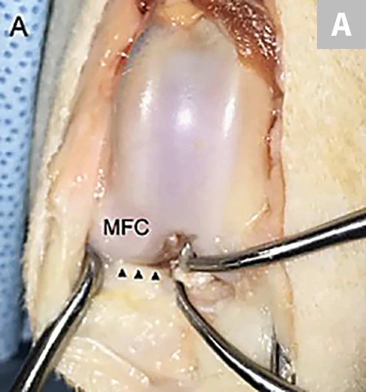
FIGURE 1 Stifle joint from a beagle viewed by arthrotomy. (A) Medial compartment view through a craniomedial arthrotomy. (B) Arthrotomy after stifle distraction using a 1.5-mm hook probe
This study investigated the diagnostic accuracy of 5 physical examination tests for predicting medial meniscal damage in cases of CCL failure. The tests, which were performed under general anesthesia, included cranial drawer (in flexion and in extension), tibial compression test (TCT), modified TCT (mTCT), and stifle range of motion (ROM). Each dog was evaluated by a novice (one of 3 interns) and by an experienced clinician (one of 4 surgeons). Meniscal tears were confirmed via craniomedial arthrotomy and probing.
Dogs with CCL failure (n = 57) were studied; 27 (47%) had concurrent tears of the medial meniscus confirmed at surgery. Of these, the types of meniscal tears included displaced vertical longitudinal (n = 18), nondisplaced vertical longitudinal (n = 7), and complex (n = 2).
The mTCT test was the most sensitive test for identifying a meniscal click by novice (63%) and experienced (59%) observers. Stifle ROM was only 44% or 33% sensitive for novice and experienced observers, respectively. The specificity of the mTCT was 77% and 73% for novice and experienced groups, respectively. Specificity of ROM was 80% for both groups. Across all tests, surgeon experience did not increase tear detection. The presence of a displaced vertical longitudinal (bucket-handle) tear was more commonly associated with a meniscal click.

These data suggest that performing the TCT under load through a ROM may improve a surgeon’s ability to preoperatively detect tears.
… The Takeaways
Key pearls to put into practice:
A meticulous orthopedic examination should be performed with the patient under sedation to assess stifle instability using cranial drawer and tibial thrust, check for presence of meniscal click during stifle manipulation, and assess for periarticular fibrosis (medial buttress) and stifle ROM.
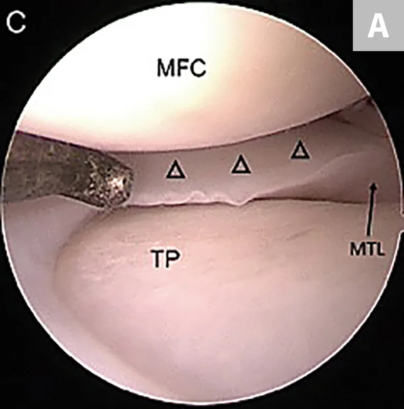
Careful joint examination is imperative during surgical treatment of CCL rupture (Figures 1 and 2). Use of stifle distractors can increase joint exposure, and probing the meniscus with a small hook probe can increase tear detection. For superior illumination and visualization, arthroscopy should be considered.
IML = intermeniscal ligament, MFC = medial femoral condyle, MTL = meniscotibial ligament, TP = tibial plateau