Image Gallery: Secondary Skin Lesions
Alexander Werner Resnick, VMD, DACVD, Animal Dermatology Center
In dermatologic diseases, abnormalities are visually apparent; thus, accurate recognition of skin lesions is key to diagnosis and management. Dermatologic diseases are often chronic, requiring clear documentation in patient medical records. Accurate recognition of skin lesions, as well as concise documentation, permits the formulation of a relevant differential list and appropriate record of disease progression and/or patient response to treatment.
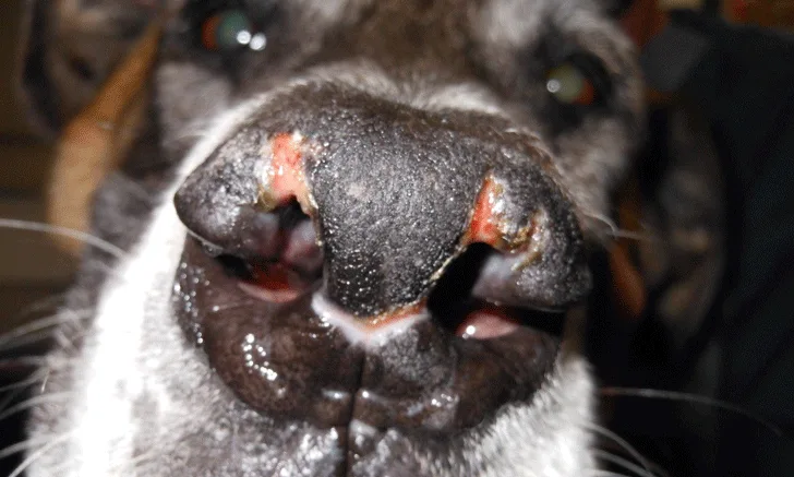
Whereas primary lesions (Part 1) involve changes in the skin caused directly by the disease process, secondary skin lesions most often develop from primary skin lesions as a result of patient or environmental factors. Several skin lesions (eg, scale, crust, stria, pigmentation change) may be considered either primary or secondary, depending on the cause. For example, hereditary abnormalities of keratinization (eg, congenital ichthyosis in golden retrievers) produce, as a primary lesion, excessive scale due to the lack of proper desquamation, with resultant accumulation of rafts of keratinocytes being shed together rather than imperceptibly as individual cells. Conversely, inflammatory dermatoses produce scale as a secondary lesion by increasing epidermal turnover, resulting in excessive production of keratinocytes that ultimately require shedding. The following images exhibit secondary dermatologic lesions.

FIGURE 1
Alopecia. Alopecia is a visible decrease in or loss of the hair coat. Secondary alopecia may result from manual removal of the hair by scratching.