Image Gallery: Primary Skin Lesions
Alexander Werner Resnick, VMD, DACVD, Animal Dermatology Center
The diagnosis of every disorder, including skin-related conditions, requires a systematic approach to a differential diagnosis list. For dermatologic diseases, abnormalities are visually apparent; thus, an understanding of basic skin lesions and their patterns of presentation can improve the differential diagnosis list and diagnostic investigation plan.
Dermatologic diseases are often chronic or recurrent. With contemporary electronic medical records, concise and correct documentation of lesions allows for more accurate recording of disease progression and/or patient response to treatment.
The dermatologic examination begins with a global review of lesion location: generalized or localized, symmetrical or asymmetrical, regional or limited to a specific structure (eg, pinnae, tail, nasal planum). Lesion localization is followed by annotation of descriptors, which may include pigmentation changes (eg, hypo- or hyperpigmentation) or hair coat qualifiers (eg, dull, brittle, thin, dry, oily). Finally, specific lesions are described to “paint a picture” with words.
Primary lesions result directly from the disease process; secondary lesions are induced from primary lesions through patient or environmental factors (some lesions may be either primary or secondary). The following image gallery provides visual presentations of various primary lesions.

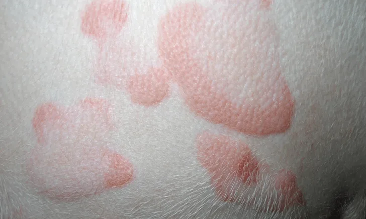
FIGURE 1
Milia. Milium are keratin-filled cysts within or just below the epidermis but without an opening to the surface. Milia are most often associated with an excess of corticosteroids (iatrogenic or natural).