Image Gallery: Canine Fundus Diseases
DJ Haeussler, Jr, DVM, MS, DACVO, The Animal Eye Institute, Cincinnati, Ohio
Fundic examinations should be performed on all patients that are presented for physical examination by the veterinarian. Abnormalities that can affect the fundus of a dog include inherited disease, acquired disease, manifestations of systemic disease, congenital abnormalities, and variations of normal anatomy. The more frequently a practitioner examines the fundus of all canine patients, the more confident he or she will become in distinguishing normal variants from abnormalities.
Click through this image gallery to see examples of a normal canine fundus and pathologies that can be easily identified on routine examination.
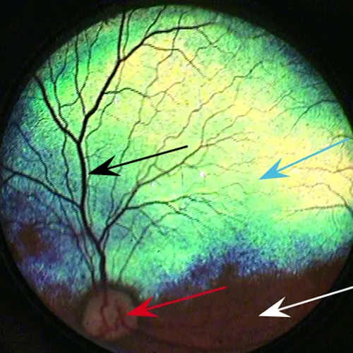
FIGURE 1 Normal canine retina
Normal canine retina in a 3-year-old spayed Chihuahua. Practitioners should be comfortable identifying the optic nerve (red arrow), tapetal fundus (blue arrow), nontapetal fundus (white arrow), and retinal vasculature (black arrow).