Identifying Causes of Otitis Externa
Jenise C. Daigle, DVM, Diplomate ACVD, Austin Veterinary Dermatology & Allergy
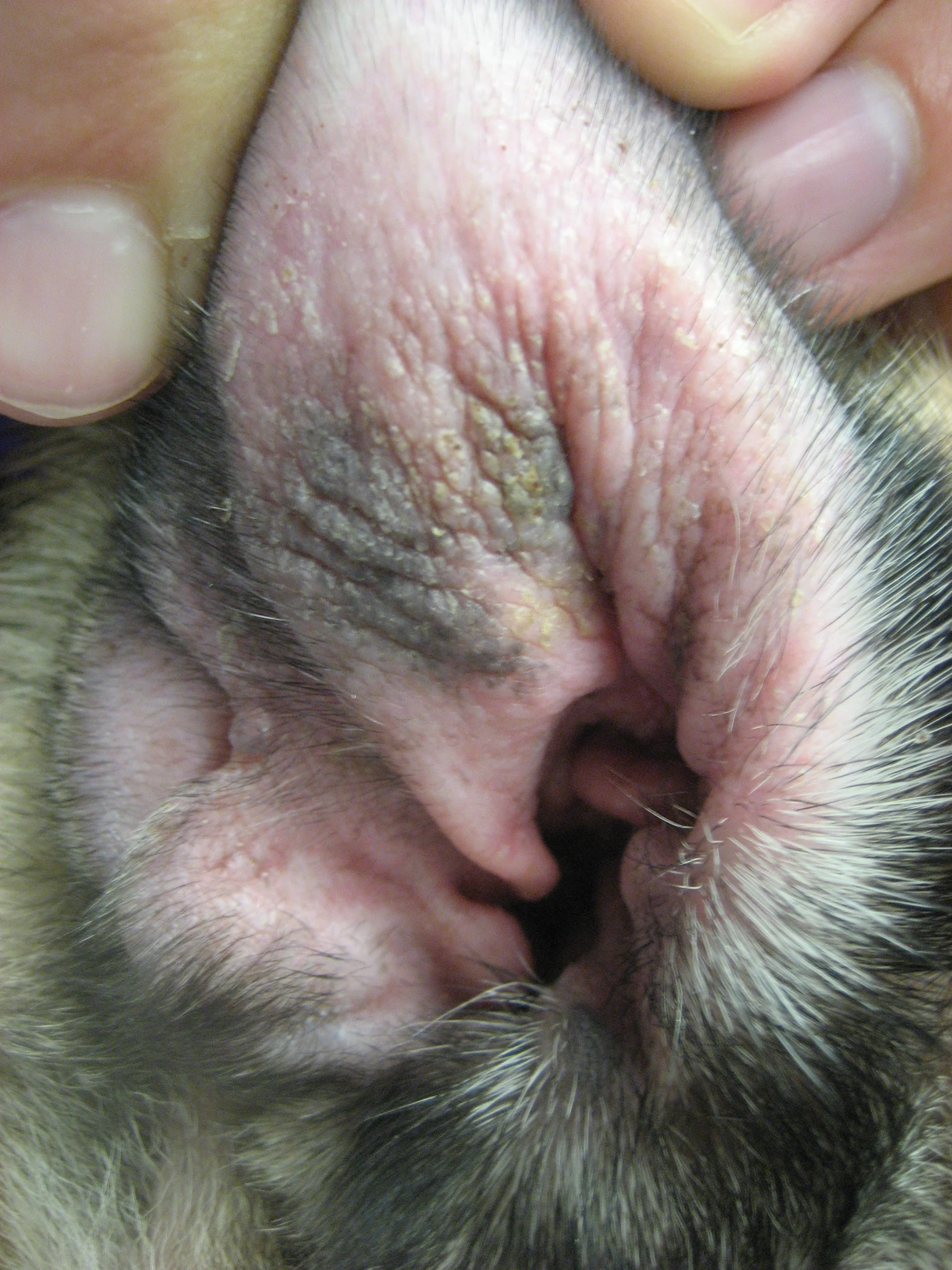
Dog with underlying allergic skin disease and concurrent allergic otitis. Note presence of erythema on the pinna with secondary lichenification and hyperpigmentation from chronic rubbing
Otitis externa is a common condition observed in everyday clinical practice.
It is usually straightforward, but successful management must involve identifying and managing the underlying cause. Causes should be classified as either predisposing, primary, or perpetuating. Recognition of these factors is critical to long term control of chronic otitis.
Predisposing Factors
The external ear canal is defined as the pinna, horizontal and vertical canals, and the tympanic membrane. Factors that directly change the microenvironment of the ear canal are predisposing factors. They facilitate inflammation by permitting the external ear canal microenvironment to be altered, which allows pathogenic or opportunistic bacteria to become established. These factors include:
Conformation of the ear canal
Pinnae (floppy vs erect ears): Evidence suggests there is a higher prevalence of otitis externa in floppy ears.
Length and conformation of ear canals
Stenosis or swelling of the opening of the external ear canal: Lack of a significant opening into the canal allows otic secretions to accumulate, providing a medium for growth of pathogenic bacteria, and may also reduce air circulation. This is a notable problem in some breeds, such as shar-peis.
Environmental factors: Heat and humidity tend to create a microenvironment that is suitable for bacterial and yeast growth. Dogs that swim may be predisposed to otitis due to excessive moisture and humidity.
Improper treatment: Trauma to the ear canal during treatment can damage the epithelial lining and predispose the canal to infections.
It is important to note that predisposing factors alone do not cause otitis; they need to be paired with another factor to cause disease. For example, a dog with floppy ears and underlying atopy is predisposed to otitis because it is easier for pathogen flora to become established.
Primary Factors
Primary factors are those that cause or initiate the inflammatory process within the ear canal. In short, they are the reason that the problem begins. Recognition and treatment of these factors is important in preventing chronic recurrent otitis.
Ectoparasites
Ear mites (Otodectes cynotis), various forms of mange (eg, sarcoptic mange, demodectic mange), and ticks- Ear mites are the most common cause of otitis externa in cats.
Allergic skin diseases (Figure 1):
These diseases are the most common cause of persistent otitis externa in dogs.- Allergies include atopy, food allergies, and contact allergies. Atopic dermatitis and food allergy should be considered more likely causes than contact allergy.
Foreign bodies
Substances in the external ear canals of dogs and cats cause irritation and inflammation. In some cases, foreign objects can damage or perforate the tympanic membrane, resulting in more serious damage.
The most common foreign bodies are plant materials (Figure 2).
Keratinization disorders: These disorders produce a ceruminous otitis externa. Primary idiopathic seborrhea and hypothyroidism may be associated with a ceruminous otitis externa.
Other dermatological diseases
Autoimmune diseases (eg, pemphigus foliaceus, discoid lupus erythematosus)
Ear tumors and polyps can cause obstruction of the external ear canal, preventing removal of normal secretions.

Piece of plant material found in the horizontal canal during lavage under anesthesia
Perpetuating (Secondary) Factors
Factors that allow inflammation and irritation to continue, even if and when the primary factor is controlled, are called perpetuating factors. These factors prevent resolution or exacerbate otitis; they must be controlled in order to prevent chronic otitis. Bacterial and yeast infections are the most common perpetuating factors.
• Bacterial infections
Most common bacteria include Staphylococcus pseudointermedius, Streptococcus species, Proteus mirabilis, Pseudomonas aeruginosa, Enterococcus species, Corynebacteria species, and Escherichia coli.
When interpreting ear cultures, remember that a low number of commensals and potential pathogens is normal. The most frequent organisms isolated in normal ears with positive cultures are Staphylococcus intermedius, coagulase-negative staphylococcus, and Micrococcus species. If these organisms are observed in low numbers on ear cytology but the animal has clinical signs (eg, erythematous pinnae/canals, head shaking, painful ears), then treatment should be considered.- Bacterial infections in the ear result in malodor, excessive production of exudate, ulceration, and extreme pain when the ear is handled.
Yeast infections
Malassezia pachydermatis is the yeast found most frequently in association with otitis externa. Malassezia organisms, in small numbers, are considered normal inhabitants of the ear canals.
Malassezia infections result in accumulation of a cream-to-dark waxy, odiferous discharge.
Otitis media (inflammation of the middle ear) may result from trauma, neoplasia of the middle ear, or, most commonly, bacterial or fungal infections. Otitis media can be a source of recurrent otitis externa.
Chronic changes
Repeated episodes of otitis externa and development of scar tissue may cause hyperplasia of the ear canal. These changes may also restrict the size of the ear canal.
Calcification of the cartilage of the ear canal results from chronic scarring and inflammation. It is irreversible and ear ablation may be the only viable treatment option.
Diagnosis
The following diagnostic tests are always useful to help diagnose causes of chronic otitis externa:
Complete medical history
Complete physical and dermatologic examination to evaluate other clinical signs of allergic skin disease
Otoscopic examination of external ear canal (Figures 3 and 4) (This may require sedation if ears are painful.)
Cytology of ear canal: A sample of exudate is taken from the ear canal, preferably at the junction of the vertical and horizontal canal, smeared onto a glass microscope slide, stained, and examined microscopically.
Necessary to identify perpetuating factors, such as bacterial or yeast infections
A MUST for follow-up examinations to determine efficacy of treatment.

Video otoscopic view of a normal ear

Ruptured tympanic membrane visualized during video otoscopy
The following tests may be helpful and necessary for proper diagnosis and treatment:
Aerobic culture of the ear canal at the level of the vertical and horizontal junction is routinely recommended. If fungal organisms are seen on cytology, then fungal culture should be requested; however, fungal otitis is rare.
Radiographs
Bulla radiographs may be indicated when otitis media is suspected.
Heavy sedation or general anesthesia is generally required.
Allergy diagnostics: Should include food trials, ectoparasite elimination, and in vitro allergy testing or intradermal skin testing. These diagnostics help identify primary factors.
When to Consider Referring
Referral should be considered if:
Otitis media is suspected and/or a myringotomy with middle ear lavage is necessary and beyond the comfort level of the practitioner
The practitioner is having trouble determining the underlying cause of chronic otitis
Neurologic signs consistent with middle or inner ear disease, such as head tilt, nystagmus, or facial paralysis, are present (aggressive medical treatment is often necessary in these cases).
If a mass is observed or suspected, referral for video otoscopic examination and surgery may be necessary (Figure 5).

Mass present in the horizontal canal of a cat
The Referral Process
When referring, it is important to provide a detailed history of previous diagnostics and treatments. It is also important to document any potential topical sensitivities the patient may have had to past treatments.
When Referral Is Not an Option
It is important to discuss with owners that compliance is an important key in the management of chronic otitis. Successful management involves identifying and managing the primary cause and treatment of inflammatory and, if present, infectious components that are causing clinical signs.
Otitis externa often requires topical corticosteroids and, in severe proliferative chronic cases, systemic therapy. In addition, dogs with chronic recurrent otitis externa should be evaluated for otitis media. Heavy sedation or general anesthesia is often required to properly examine the external ear canal and middle ear.
Owners also need to understand the importance of follow-up evaluations. These examinations, which document resolution of clinical disease, not just resolution of clinical signs, are a must.