Icterus & Collapse in a Cavalier King Charles Spaniel
Leslie Sharkey, DVM, PhD, DACVP (Clinical Pathology), Cummings School of Veterinary Medicine at Tufts University
A 7-year-old neutered male Cavalier King Charles spaniel is presented after an episode of collapse.
History
The dog was presented to the referring veterinarian after an episode of collapse and weakness at a boarding kennel. The patient had been eating and drinking normally before the collapse episode but had been lethargic. The referring veterinarian noted evidence of hemolytic anemia, which was suspected to be immune-mediated. The dog was referred after initial treatment with vitamin K, gastroprotectants, and IV fluids. An untreated heart murmur was noted on the history.
Physical Examination
On presentation to the referral hospital, the patient had icteric sclera and mucous membranes, a grade 3/6 systolic heart murmur, discomfort on abdominal palpation, nystagmus, and decreased pupillary light responses and mentation.
Laboratory Results
A complete blood count revealed marked neutrophilic leukocytosis with a moderate regenerative left shift, including rare metamyelocytes, toxic change, and monocytosis. Other laboratory findings included a hematocrit of 9%, mild macrocytosis, normal mean corpuscular hemoglobin concentration, increased nucleated red blood cells, and normal platelet count. A review of the blood film revealed 50% to 75% Heinz bodies, numerous spherocytes, and red blood cell ghosts (Figure 1).

A. Normal canine red blood cells; B. large Heinz body (arrow); C. spherocyte (note the absence of central pallor compared with normal red blood cells); D. ghost cell with retained Heinz body (arrow)
Ask Yourself
What potential causes of hemolytic anemia would be consistent with all of the morphologic abnormalities in the erythrocytes?
What further diagnostic tests are recommended to determine the cause of the hemolysis in this patient?
Diagnosis
Probable zinc toxicity
Other differential diagnoses could include ingestion of other oxidants, such as acetaminophen, onions, or garlic.
Additional Diagnostics
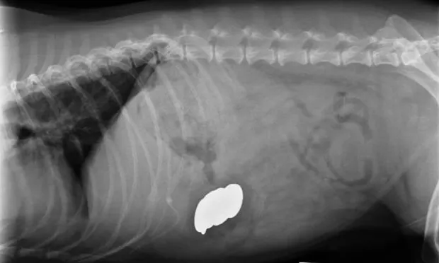
Abdominal radiography revealed metallic foreign bodies (Figure 2) in the stomach. Multiple coins, including some zinc containing pennies minted after 1982, and a medallion were removed endoscopically (Figure 3). Blood zinc levels were not measured.

Lateral abdominal radiograph revealing metallic foreign bodies
Zinc may directly inhibit coagulation factors in the intrinsic pathway, resulting in prolonged activated partial thromboplastin time with minimal changes in the prothrombin time. Secondary disseminated intravascular coagulation is also possible. In this dog, the prothrombin time was mildly prolonged at 9.2 seconds (normal range, 6.2–7.7 seconds) and the activated partial thromboplastin time was moderately prolonged at 39.5 seconds (normal range, 9.8–14.6 seconds).
Serum biochemical profile abnormalities included azotemia, which may have been prerenal or attributable to zinc-induced renal tubular necrosis, and hyperbilirubinemia with moderately increased alkaline phosphatase and markedly increased alanine and aspartate aminotransferase levels. Liver enzyme abnormalities are very common in dogs with zinc toxicity, and they may be due to direct toxic hepatocellular damage or hemolysis, resulting in hepatic tissue hypoxia. The plasma was also markedly hemolyzed, consistent with an acute hemolytic event.

Endoscopically removed foreign bodies; note the corroded appearance of the pennies.
Treatment
Medical reduction of gastric acid production with histamine-2 receptor antagonists or proton-pump inhibitors may decrease zinc absorption until zinc containing foreign bodies can be removed surgically or endoscopically. Induction of vomiting for removal may not be successful and could worsen mucosal damage to the esophagus and stomach. Chelation therapy has been reported, but no systematic evaluation of benefits has been published in the veterinary medical literature.
Initial stabilization of this patient included fluid therapy to correct dehydration and induce diuresis, and blood products (packed red blood cells and plasma) to ameliorate tissue hypoxia and coagulation abnormalities. The patient also received gastroprotectants.
Outcome. Despite aggressive supportive therapy and removal of the zinc, the patient experienced cardiac and respiratory arrest. Necropsy suggested that disseminated intravascular coagulation was the likely cause of death.
Ask Yourself
1. What potential causes of hemolytic anemia would be consistent with all of the morphologic abnormalities in the erythrocytes?
Causes of Heinz body anemia in the dog include exposure to zinc, Allium species (garlic and onion), vitamin K3, and acetaminophen. Causes of spherocytes include immune-mediated damage to erythrocytes, zinc toxicity, and bee and snake envenomation. A few spherocytes may be associated with any cause of Heinz body anemia; however, when large numbers of spherocytes and Heinz bodies are present simultaneously, zinc toxicity is an important differential diagnosis. Heinz bodies are considered the primary indicator because spherocytes are present in only a minority of cases.
2. What further diagnostic tests are recommended to determine the cause of the hemolysis in this patient?
Further diagnostic tests should include chest and abdominal radiography to evaluate the patient for the presence of metallic foreign bodies in the esophagus, stomach, or gastrointestinal tract, along with evaluation of blood zinc levels. The result of a Coombs test may be positive in patients with zinc toxicity, and it does not distinguish this condition from idiopathic immune-mediated hemolytic anemia.