Heterobilharzia americana Infection & Schistosomiasis in Dogs
Nicole Szafranski, DVM, University of Tennessee
Richard Gerhold, DVM, MS, PhD, DACVM, University of Tennessee
In the Literature
Graham AM, Davenport A, Moshnikova VS, et al. Heterobilharzia americana infection in dogs: a retrospective study of 60 cases (2010-2019). J Vet Intern Med. 2021;35(3):1361-1367.
The Research …
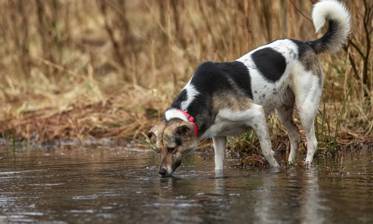
Heterobilharzia americana is the causative agent of canine schistosomiasis. Although raccoons are the natural host, dogs can act as definitive hosts. Canine schistosomiasis has historically been diagnosed in the Gulf Coast of the United States, but recent studies report an expansion in geographic range as far as Kansas and Utah.<sup1,2 sup>
The free-swimming life stage (ie, cercaria) of the parasite leaves its intermediate host, an aquatic lymnaeid snail, and penetrates the skin of a raccoon or dog. From the skin, it migrates through the lungs and liver to the mesenteric veins to mate. Eggs are deposited in the host’s intestinal lumen and passed in the feces. Hematogenous dissemination of eggs causes severe granulomatous reactions in dogs. Typically, young large-breed dogs have a greater risk for exposure, but any dog that comes in contact with potentially contaminated water is at risk.
H americana generally causes granulomatous GI and hepatic disease; clinical presentation and signs can vary.3 Diagnosis can be challenging and may require submission of feces to a diagnostic veterinary parasitology laboratory. Saline fecal sedimentation and/or histopathology have been the primary diagnostic tools used to diagnosis H americana. A sensitive fecal PCR test was recently developed and can help in the earlier diagnosis of, and potential improved prognosis for, H americana infection.4
This retrospective study examined the medical records of 60 dogs from 2 veterinary referral hospitals in Texas from 2010 to 2019 to explore clinical findings, diagnostic methodology, and treatment outcomes. The most common clinical signs included diarrhea (55.8%), vomiting (46.2%), and weight loss with or without anorexia (15.4%). Laboratory abnormalities included hyperglobulinemia (42.6%), elevated liver enzymes (30%), moderate eosinophilia of >500/µL (42.3%), anemia (17.6%), and hypercalcemia (11.6%). Ultrasound images disclosed pinpoint hyperechoic foci in the intestines, liver, or mesenteric lymph nodes in 64.4% of dogs.
Although diagnostic methods varied between patients, 81.7% of dogs were diagnosed with H americana via PCR alone. Of the remaining dogs, 6 were diagnosed via biopsy, 3 via either direct fecal smear or sedimentation, and 2 at necropsy. Treatment plans were highly individualized, but the majority included a combination treatment of praziquantel and fenbendazole, with retreatment as needed on a case-by-case basis. Survival information was available for 34 of 56 dogs that underwent treatment; 25 of the 34 dogs (73.5%) were still alive 6 months after diagnosis. Mortality rate was thus 17.6%.
… The Takeaways
Key pearls to put into practice:
H americana infection should be considered in dogs with chronic GI signs and a history of exposure to outdoor water sources.
Ultrasonography and fecal PCR should be considered as standard testing methods to aid in earlier diagnosis of H americana and potentially provide better treatment outcomes.
Increased awareness of H americana is warranted because of its geographic spread.
You are reading 2-Minute Takeaways, a research summary resource presented by Clinician’s Brief. Clinician’s Brief does not conduct primary research.