Hepatobiliary Imaging With Radiography & Ultrasonography: Biliary System
Lorrie Gaschen, DVM, PhD, Dr.med.vet, DECVDI, Louisiana State University
Abdominal radiography is important in the diagnosis of hepatobiliary disease, as it can help detect abnormal liver size and shape and identify mineralization or gas in the liver or intra- and extrahepatic biliary tract. Abdominal ultrasonography is the most useful noninvasive diagnostic tool for differentiating hepatic from posthepatic causes of icterus, obstructive from nonobstructive biliary disease, and diffuse from focal hepatic parenchymal disease.1
This article is part of an illustrative series on hepatobiliary imaging, including both the liver and biliary system.
The liver, gallbladder, biliary tract, duodenal papilla, and pancreas should be examined with a high-frequency transducer, and, because these structures are in the cranial abdomen, it is usually best to use a curved-array transducer with a small footprint.
Ultrasonography is useful for estimating hepatic size and evaluating internal architecture (including portal, venous, arterial, and biliary vasculature; echogenicity; and echotexture). Size, wall thickness, and contents of the gallbladder, as well as size of the bile ducts, can also be assessed. Ultrasonography is also helpful for differentiating chronic injury (eg, cirrhosis, end-stage chronic inflammatory disease) from acute liver injury based on the size, shape, and internal architecture of the liver parenchyma.2
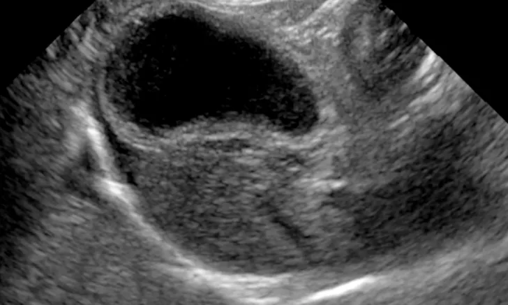
The gallbladder wall and contents can be assessed for cholecystitis, cholelithiasis, and mucoceles via ultrasound. Ultrasound-guided tissue sampling of the liver parenchyma and cholecystocentesis is safe and often necessary due to the nonspecific nature of imaging findings.3-6

FIGURE 1A
Lateral radiographs of a clinically normal cat (A) and a 14-year-old neutered male domestic shorthair cat (B) presented with icterus and elevated liver enzymes. A normal feline liver should have a sharp, pointed margin (A; arrows) and should not extend past the costal arch; the gastric axis should be parallel to the ribs (A; line). The liver in the icteric cat extends past the costal arch, and has rounded borders (B; arrows). The stomach is displaced caudally (B; line; gastric axis from the fundus to the antrum). An ovoid mineralization (B; arrowheads) is also superimposed with the cranioventral liver in the location of the gallbladder. This cat was diagnosed with cholangitis, lipidosis, and a cholelith.
This article is part of an illustrative series on hepatobiliary imaging. See the companion article for images of the liver.