Hairline Bone Fracture
Hairline fractures are uncommonly diagnosed in small animal patients, but may occur more frequently than suspected. These fractures may cause lameness or pain on palpation. For the purposes of this article, a hairline fracture is defined as any disruption of normal bone integrity that is not clinically obvious on radiographs. Small fractures may be difficult to see on survey radiographs due to minimal displacement of the fracture fragments, fracture of one cortex only, or presence of superimposed structures. Hairline fractures may occur in any type of bone and can be intraarticular. They may be caused by a single traumatic incident or by repetitive trauma (stress fracture). Stress fractures are often unicortical and difficult to diagnose. Intraarticular hairline fractures may propagate and eventually displace, becoming chip fractures. Some hairline fractures may actually result from developmental failure to ossify rather than being a true fracture.
Diagnosis of hairline fractures can be assisted by alternative radiographic positioning, oblique and skyline radiographic views, serial radiographic examination, nuclear scintigraphy (bone scan), CT, MRI, arthroscopy, or exploratory surgery. Traditional radiographic equipment can be used to make a diagnosis of a hairline fracture in most patients. Oblique and skyline views are particularly useful because they displace superimposed structures, allowing an unobscured view of a particular area of the bone. Serial radiographic examination every 4 weeks is also useful because hairline fractures will often become more evident over time due to bone resorption and periosteal proliferation.

Figure 1A. Hairline fracture of the fibula.

Figure 1B. The fracture is more clearly seen 4 weeks later following bone resorption and periosteal proliferation at the fracture site.

Figure 2A. The capital physis of this dog, which presented with hip pain, appears slightly widened.

Figure 2B. If the patient is positioned in a frog-legged position, the hairline fracture displaces and is easily diagnosed.
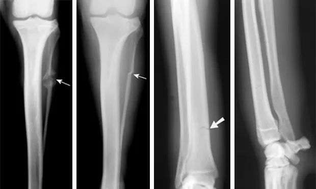
Figure 3A. This series represents an example of a probable fracture that cannot be seen on routine radiographs but that produces secondary signs that can be seen. Slight thickening of the caudal cortex of the tibia (arrow) is present in a dog with left hind limb lameness. The dog is an active, 3-yearold, castrated male black Labrador retriever having lameness for several months but with no prior history of lameness.

Figure 3B. Lameness was persistent, and cortical thickness progressed (arrow) over a 4-month period. Panosteitis might be considered in the differential diagnosis because of the several areas of uptake, although panosteitis does not typically start as a focal unicortical thickening of the bone in a 3-year-old dog.

Figure 3C. A bone scan was positive in the shaft of the tibia and was suggestive of a hairline stress fracture.

Figure 3D. Several small holes were drilled in the affected cortex (osteostixis) to encourage neovascularization and bone healing. The lameness resolved after treatment.
Figure 4A. A 5-year-old, male Brittany spaniel with a 2-month history of lameness. The opposite elbow was normal. A fracture line cannot be seen in this anteroposterior view of the elbow.

Figure 4B. A hairline intercondylar fracture (arrow) can be seen after repositioning the elbow and x-ray beam only slightly. This may represent incomplete ossification or a true fracture. In this case, the hairline fracture was confirmed by bone scan and arthroscopy.

Figure 5A. An obvious fracture is not seen on this lateral radiographic view of the radius and ulna.

Figure 5B. A nondisplaced hairline fracture of the distal radius (arrow) that involves the medial cortex of the distal radius is easily seen, demonstrating the need for multiple radiographic views.
AcknowledgmentThe author thanks Drs. Brian Poteet, Michelle Fabiani, Bill Liska, and Anne Bahr for providing case material.