Grinning Puppy
History. A 4-month-old, intact female German shepherd presented for inability to open the jaw. The owner reported an alteration in ear carriage 2 days before presentation. The puppy had also been lethargic and anorectic for approximately 24 hours. Routine vaccinations and deworming were up-to-date, and there was no history of previous illness, exposure to toxins, or interaction with other animals.
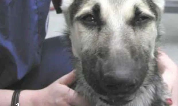
Examination. The patient was lethargic with a rectal temperature of 104° F. The heart and lungs were normal on auscultation, although the respiratory rate was elevated at 60 breaths/min. The jaw was unable to be opened, and ptyalism was profound. The ears and muscles of facial expression were severely contracted (Figure 1). The results of the remainder of the physical and neurologic examinations were within normal limits.
Ask Yourself ...
What is the most likely diagnosis for this 4-month-old puppy?A. Congenital myotoniaB. TetanusC. Chronic masticatory myositisD. Strychnine poisoningE. Temporomandibular disease
Correct Answer: B
Tetanus, which is caused by the organism Clostridium tetani, is the most likely disorder in this patient.
The medical term for inability to open the jaw is trismus (lock jaw). Trismus is defined as a firm closing of the jaw due to tonic spasm of the muscles of mastication. It is most commonly seen with both the focal and generalized forms of tetanus, but inability to open the jaw may also occur with mechanical instability associated with such conditions as chronic masticatory myositis and temporomandibular disease.
The descriptive term for the abnormalities in this dog's facial muscles and pinna is risus sardonicus-the semblance of a grin caused by facial spasm, usually in tetanus.
Diagnostics. Serum antibody titers to tetanus toxin can be measured for confirmation, but clinical signs and physical examination alone are generally sufficient to establish a diagnosis. A recent history of a wound or the presence of a wound is common, although isolation of the organism is difficult and often unrewarding. The disorder may be seen postsurgically, but in teething puppies the source of infection is most commonly the oral cavity; therefore, clinical signs are initially observed in the head. In this case, two infected teeth were identified and removed during an oral examination (Figure 2). No other wounds were noted in this animal.
Treatment. Management of severely affected animals is costly, and the clinical course can be protracted, requiring weeks of hospitalization. Initial treatment includes IV administration of antitoxin (100 to 1000 U/kg) and aggressive wound management. Due to the high potential for anaphylaxis after IV antitoxin, animals should be premedicated with antihistamine. An intradermal test injection of 0.1 to 2 ml should be done approximately 15 to 30 minutes before the antitoxin is administered, and the site must be observed for wheal formation. Antibiotics, such as penicillin G or metronidazole, should also be given, although they will not be effective against circulating toxin.
Supportive care. Supportive care is key and includes muscle relaxants and sedatives (e.g., diazepam, acepromazine, methocarbamol, and barbiturates), soft bedding, a dark and quiet environment, urinary catheterization when needed, and nutritional support. A feeding tube is frequently required because of the potential for masticatory and pharyngeal dysfunction and the high incidence of aspiration and death.
In the patient described here, the clinical signs began to worsen 12 hours following presentation and generalized tetanus was apparent. Treatment measures included antitoxin administration, IV diazepam and acepromazine, metronidazole, histamine blockers, metoclopramide, and placement of a percutaneous endoscopic gastrostomy tube. Fortunately, this animal made a full recovery.
Take-home message
Trismus is one of the most common clinical signs of tetanus, which must be distinguished from chronic masticatory myositis. The absence of recent surgery or a wound does not rule out tetanus.
TX ... at a glance
• Intradermal antitoxin test injection, 0.1 - 2 mlObserve for wheal formation; then 15-30 min later:• Premedicate with histamine blockers• Antitoxin, 100 - 1000 U/kg IV• Antibiotics (e.g., penicillin G, metronidazole)• Supportive care:TM Muscle relaxants and sedatives (e.g., IV diazepam and acepromazine)TM Soft beddingTM Dark, quiet environmentTM Urinary catheter as neededTM Feeding tube as needed