Video capsule endoscopy (VCE) involves generation of images by telemetric recording of the luminal GI tract via an ingestible capsule that contains cameras and spontaneously travels through the GI tract.1,2 VCE is safe and well tolerated and can help diagnose the origin of GI bleeding in dogs with careful patient selection but should not be used in patients with bowel obstruction (eg, stenosis).3-6 In dogs with overt GI bleeding, 77% of VCE examinations showed positive findings.5 The most commonly used capsule is 11 × 31 mm and therefore only recommended for administration in dogs >9.9 lb (4.5 kg). Dogs are typically fasted 16 hours before and 8 hours after video capsule administration. Pet owners are often given a preaddressed mailing envelope and instructed to send the excreted capsule to a specialist for image analysis.
Advantages of Video Capsule Endoscopy
VCE has several advantages compared with traditional GI endoscopy, including visualization of the entire small intestine without sedation or general anesthesia (traditional endoscopy does not reach the distal duodenum, jejunum, or colon). The main indications for VCE in humans and dogs include overt or suspected occult GI bleeding in a patient with normal results on traditional endoscopy. Irregular mucosa, ulceration, abnormal vessels, dilated lacteals, masses, and bleeding lesions can be detected with VCE in dogs.4-6
Disadvantages of Video Capsule Endoscopy
Disadvantages of VCE include inability to acquire biopsy samples, uncontrolled capsule movement, and absence of insufflation.7 Incomplete small bowel studies (capsule does not reach the colon during recording time) are the most commonly reported complication in humans (20%-30%) and dogs (6%-38%).5,8 The difference in completion rates may be due to patient health status (eg, healthy vs sick, ambulatory vs nonambulatory, acute vs chronic disease), patient size, video capsule properties (eg, size, battery life), and/or preparation protocol (eg, fasting time, polyethylene glycol administration). Localizing the capsule in a segment of the small intestine can be difficult; however, landmarks (eg, duodenal papilla, ileocolic junction, transit time) can help estimate the location in the proximal, mid, or distal segment. The capsule may be retained in the stomach up to 1 week, but gastric retention (>2 weeks) has not been reported in dogs.5 Prokinetic administration or emesis induction can recover the capsule.
Video Capsule Endoscopy Images
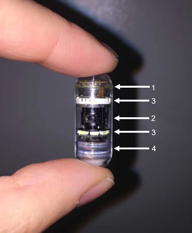
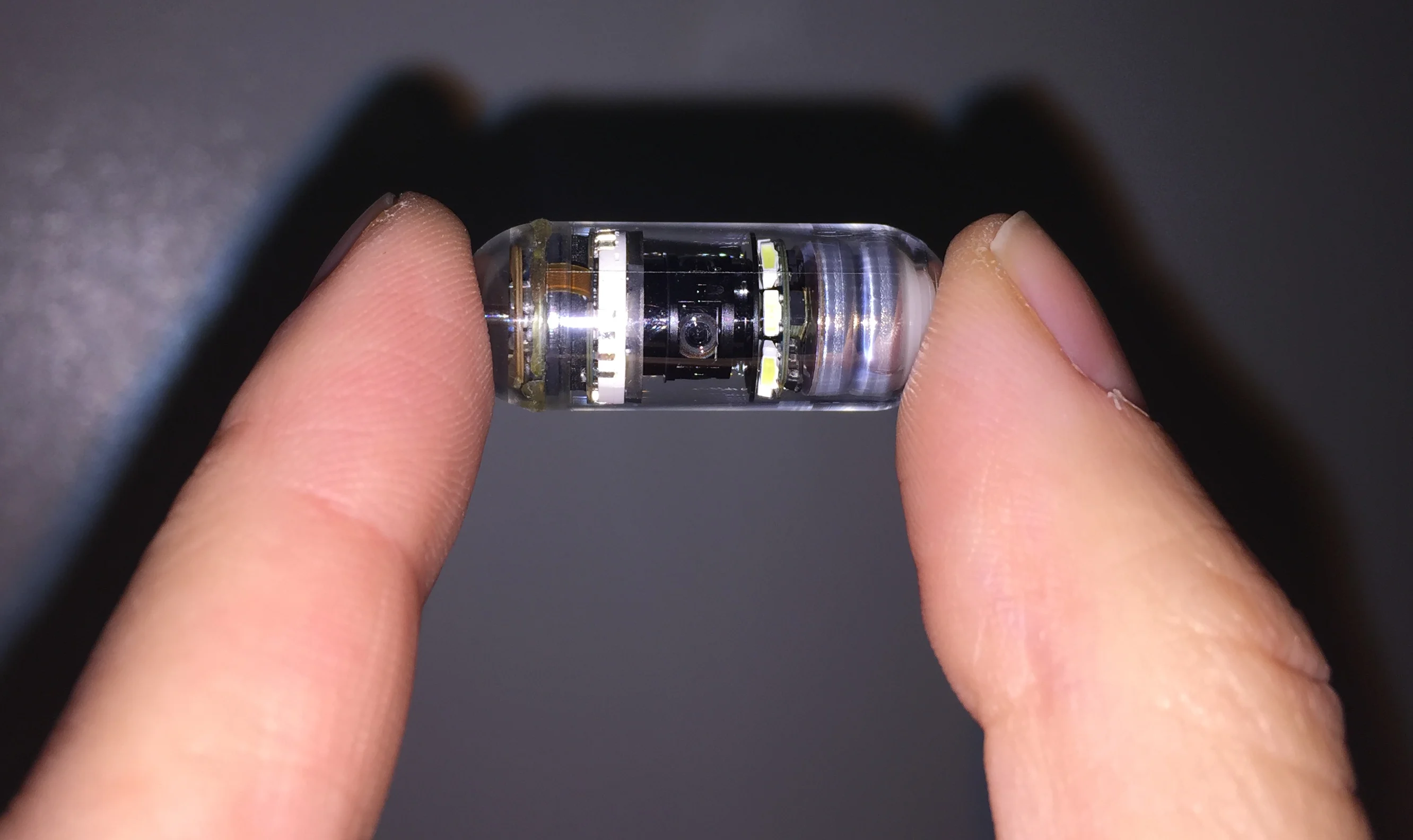
Capsules with lateral viewing (several cameras at the center of the capsule) are most widely used in veterinary medicine because of practicality (no data recorder needed) and organized interpretation by specialists. Capsules are single-use devices that can include a microprocessor and internal memory system (1), autofocusing cameras (2), LED lights (3), and batteries (4). Cameras can capture up to 5 frames per second, and battery life can range from 15 to 20 hours.
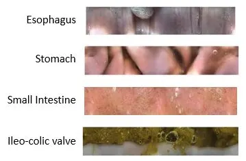
Appearance of landmarks in the esophagus, stomach, small intestine, and colonic mucosa are used by specialists when reviewing video capsule recordings.
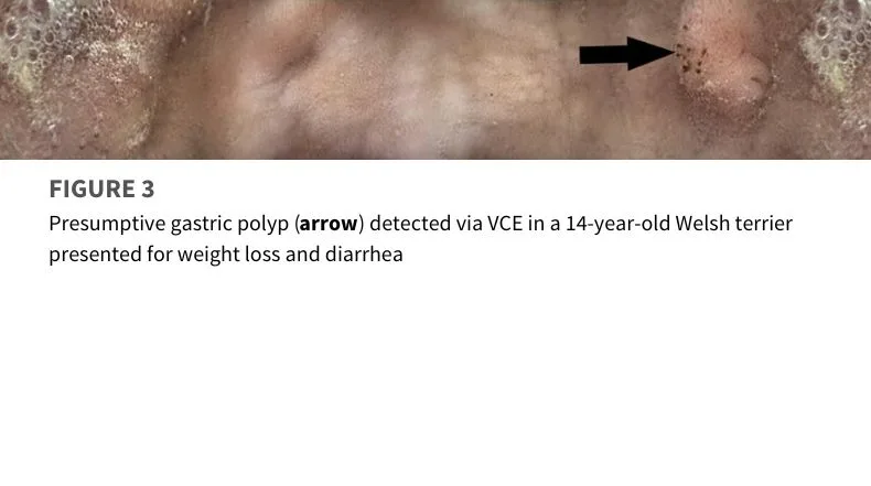
Case Studies