GI Disease in Ferrets
Angela M. Lennox, DVM, DABVP, Avian & Exotic Animal Clinic, Indianapolis, Indiana
Profile
The ferret is a laboratory model for research on emesis, Helicobacter mustelae gastritis, and certain GI neoplasms.
GI disease, common in pet ferrets, is often multifactorial and can be secondary to other disease processes and stress.
The ferret’s relatively short GI tract predisposes it to GI disease.
Causes include primary GI disease (eg, infectious, neoplastic, foreign body) and inflammatory conditions (Table: Types of GI Disease in Ferrets).
Viruses frequently affecting the GI tract include ferret enteric coronavirus (FRECV) (also known as epizootic catarrhal enteritis [ECE] virus) and ferret systemic coronavirus (FRSCV), a newly recognized virus.
In an informal survey at the author’s clinic, owners reported mild to severe diarrhea after a traumatic event (eg, change in social structure, housing, veterinary visit).
Types of GI Disease in Ferrets
Canine distemper virus
Salmonellosis
Influenza virus
Mycobacteriosis
Campylobacteriosis
| | Foreign body |
Younger ferrets most affected
Documented foreign materials include foam, rubber, and plastic
Trichobezoars are also seen
| | Infectious disease |
Bacterial, including H mustelae
Viral, including enteric (FRECV) or systemic coronavirus (FRSCV) or rotavirus in very young animals
Parasitic, most commonly coccidia in younger animals
| | Inflammatory disease |
Inflammatory bowel disease, eosinophilic gastroenteritis
Underlying causes uncertain
| | Neoplastic disease |
Primary GI neoplasms, including lymphoma and carcinoma
|
Stress from any other disease processes, including non–GI neoplasia, organ failure
| | Psychological stress |
Changes in housing or social structure, separation, veterinary visits
|
Physical examination and historical findings can reveal:
Weight loss
Decreased appetite to hyporexia or anorexia
Lethargy
Vomiting and regurgitation
Abnormal stool (eg, diarrhea, hematochezia)
Palpable masses or bowel thickening
Apparent pain on abdominal palpation
Dehydration
Emaciation and hypovolemia (in severe cases)
Rectal or colon prolapse (young ferrets) or straining from sacculectomy complications

Safe restraint of a ferret using scruff technique
Diagnosis
Definitive diagnosis requires identifying underlying primary disease, other concurrent disease process(es), and/or stress.
Primary pathogen testing includes bacterial culture and sensitivity testing, PCR assay for specific pathogens (eg, viruses), and fecal flotation (Table: Resources for Specialized Diagnostic Tests).
Fecal flotation may identify coccidia or other organisms, but GI parasitism is uncommon in pet ferrets.
Confirmation of disease from H mustelae requires gastric biopsy with documentation of lesion-associated organisms. Because most pet ferrets are thought to harbor this organism, PCR assay with biopsy is recommended.
Occult fecal blood testing can be a useful adjunct test in identifying GI hemorrhage.
Resources for Specialized Diagnostic Tests
| Cryptosporidium sppHelicobacter sppMycobacterium spp | Research Associates Laboratory | | FRECV/FRSCVCanine distemper virus | Michigan State University | | FRECVCryptosporidium sppMycobacterium sppH mustelaeCampylobacter jejuniLawsonia intracellularis | Veterinary Molecular Diagnostics |
Abdominal radiography is most useful in cases of GI obstruction and can also identify other abnormalities.
Abdominal ultrasonography can be useful, especially for investigation of masses potentially related to the GI tract.
GI tract biopsy is indicated in patients that are unresponsive to therapy.
CBC and serum biochemistry profile analysis can provide useful information on overall clinical condition (eg, hypoproteinemia from albumin loss via the GI tract).
CBC can also help identify other underlying disease conditions.
Treatment
Treatment includes resolution of fluid deficits (eg, shock, anemia, dehydration).
Blood transfusion can be relatively easy in ferrets, which have no identifiable blood types.
Some GI diseases—particularly gastritis—appear particularly painful.
Analgesics or proton pump inhibitors may be useful; the author finds famotidine to be efficacious.
Bacterial GI disease is treated with appropriate antimicrobials based on culture and sensitivity test results if available.
Good empirical choices include amoxicillin and enrofloxacin.
Treatment of viral disease is supportive only.
A regimen for FRSCV has been proposed and is based on treatment for feline infectious peritonitis (Suggested Treatment Protocols for FRSCV).
Suggested Treatment Protocols for FRSCV1*
In combination:
Doxycycline at 10 mg/kg PO q12h
Cimetidine at 10 mg/kg PO q12h
Polyprenyl at 3 mg/kg PO 3× weekly
Pentoxifylline at 20 mg/kg PO q12h
Feline vitamin supplement
Prednisolone at 1–2 mg/kg PO q12h, tapering dosage
*Currently proposed for management of ferrets with FRSCV; treatment may be long-term, depending on response to therapy
Triple therapy (metronidazole–amoxicillin–bismuth subsalicylate) is often used for suspected Helicobacter spp gastritis; other drug combinations have also been used.
Foreign body obstruction requires laparotomy. Ferrets are generally good surgical candidates, excluding those with chronic disease and marked debilitation.
A number of protocols have been developed for treatment of GI lymphoma. Success can vary.
Many ferrets with GI disease require hand-feeding.
Ideal products include high-protein, easily digestible foods that can be fed by syringe (Oxbow Carnivore Care, Hill’s Prescription Diet a/d ).
Stress, a contributor to enteritis and diarrhea, should be managed.
Ferrets that are unresponsive to therapy should be referred for advanced diagnostic testing and therapy.
Many owners can aid in treatment by administering medications and providing meals via frequent hand-feeding.

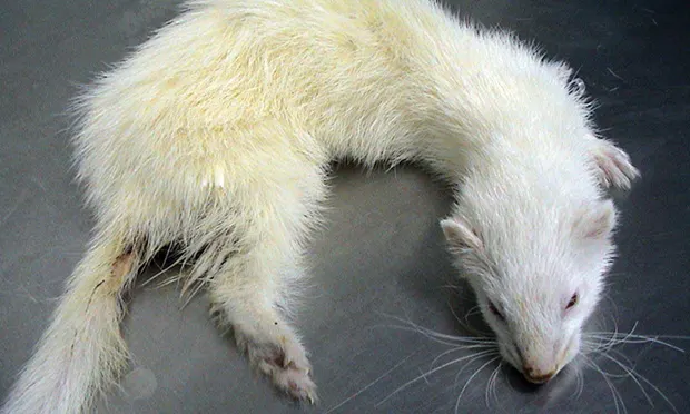
An emaciated, wasted ferret with chronic diarrhea; note dark tarry stool accumulation at the perineum, likely indicating hematochezia.
Follow-up & Prognosis
Patients must be monitored carefully for response to therapy; simple bacterial gastroenteritis may resolve promptly.
Many conditions, however, may only temporarily respond or not respond at all.
Prognosis is good for patients with bacterial or parasitic disease.
Chronic inflammatory disease may be difficult to treat.
FRECV carries a high morbidity, but most ferrets survive with supportive care.
Most deaths are seen in older or already debilitated animals.
Prognosis is guarded for ferrets affected with FRSCV.
GI neoplasia often carries a guarded prognosis; lymphoma may respond to chemotherapy.
Failure to respond is often a result of incomplete diagnosis or failure to manage other concurrent disease or stress.
Coronavirus may produce significant damage to the intestinal mucosa; chronic GI dysfunction is common and may be difficult to resolve.
ECE = epizootic catarrhal enteritis, FRECV = ferret enteric coronavirus, FRSCV = ferret systemic coronavirus