Gastrointestinal Perforation
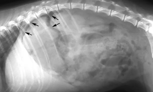
Figure 1. Crescent and triangular areas of free intraperitoneal gas (arrows) are shown that do not correspond to the tubular-shaped pattern of gas seen in the small intestine.(A) Lateral view, above. (B) Ventrodorsal view

Related Article: Surgical Treatment for Bite Wounds
The two most common causes of free intraperitoneal gas are external perforating wounds (including those caused by exploratory surgery) and bowel perforation. A large volume of gas is readily detectable on survey radiographs because the contrast of free intraperitoneal gas leads to improved visualization of serosal surfaces. Smaller amounts of gas may be more difficult to detect and may resemble bowel gas. Gas may accumulate between the liver and diaphragm, in which case the abdominal surface of the diaphragm is visualized. Because gas rises to the highest portion of the abdominal cavity, positional radiography may aid in diagnosis. This may be accomplished by placing the patient in left lateral recumbency and directing the beam ventrodorsally (horizontal beam). Gas usually localizes under the highest portion of the right abdominal wall under the caudal ribs, extending caudally under the diaphragm or abdominal wall. Gas may not collect under the highest point if trapped between viscera-a negative finding still does not eliminate free gas. A barium contrast study is contraindicated in suspected cases of bowel perforation.

Figure 2. Horizontal-beam image of the same patient as in Figure 1. A collection of gas underneath the uppermost ribs and diaphragm can be seen. At surgery, the dog had a bowel perforation secondary to a foreign body.

Figure 3. Thorax of a cat with intestinal adenocarcinoma(lateral view). Note the gas opacity caudal to the diaphragm (black arrows) that shows a separation of the diaphragm and liver (white arrow).

Figure 4. Triangular-shaped gas opacities underneath the caudal ribs and extending along the abdominal wall (horizontal-beam image). Erosion of the intestinal adenocarcinoma with subsequent perforation was noted at surgery.