Fine-Needle Aspiration of the Liver
Daniel VanderHart, DVM, DACVR
Clifford R. Berry, DVM, DACVR, University of Florida
Fine-Needle Aspiration
Liver cytology via fine-needle aspiration (FNA) has been shown effective in diagnosing some liver disorders; however, cytologic examination of the liver does have limitations and must be interpreted within the context of the clinical picture.1-5
FNA is a relatively low-risk procedure that usually can be performed without sedation or general anesthesia. Contraindications for liver FNA include coagulopathies, lack of a safe access route (eg, vascular structure in the biopsy path), inexperience on the part of the sonographer, and an uncooperative patient.
Related Article: Liver Ultrasound-Guided Fine-Needle Aspiration
Related Article: Top 5 Liver Conditions in Dogs
Step-by-Step: Ultrasound-Guided Fine-Needle Aspiration Cytology of the Liver
What You Will Need
25-or 22-gauge, 1.5-inch needles
6-mL syringe
Microscope slides labeled with patient name and tissue source
Usually, 25-gauge needles are a good choice with regard to sample size, minimizing hemodilution, and decreasing patient discomfort. Because some lesions do not exfoliate well, a larger-gauge needle can be used.
A syringe filled with room air is used to blow the sample onto the microscope slide.


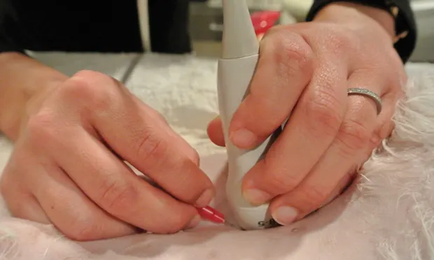
STEP 1
Prepare the area. If the patient is not already clipped, clip and clean the sampling area; consider a surgical scrub if the skin is particularly dirty or a sample will be submitted for culture. Remove ultrasound gel applied to the area, as it can create an artifact on the slide to be viewed by the pathologist.
Leave the area wet with alcohol or liquid (not jelly) lidocaine as a contact medium for the ultrasound transducer. Clean the transducer by placing an alcohol gauze sponge on the transducer tip while preparing the patient.