Feline Heartworm Infection
Clarke E. Atkins, DVM, Diplomate ACVIM (Internal Medicine/Cardiology), North Carolina State University
In addition to serologic tests, thoracic radiography and cardiac ultrasonography are important diagnostic tests in feline heartworm infection.
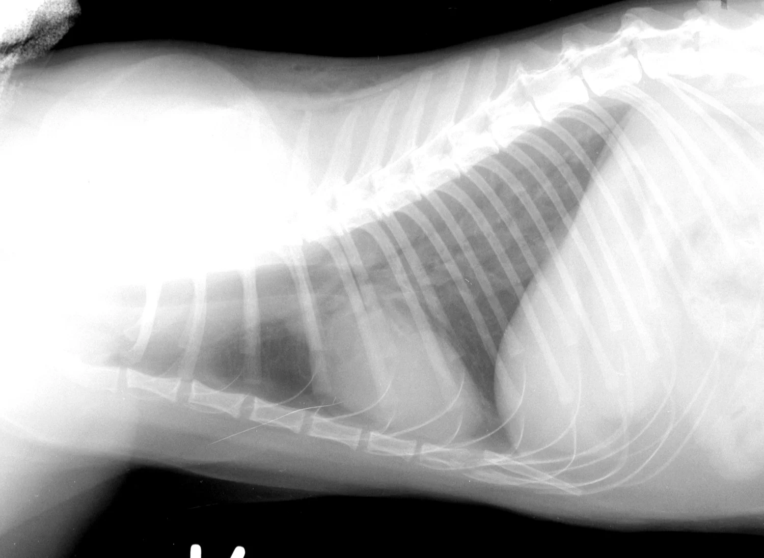
The cardiac silhouette of a cat with heartworms rarely has the "inverse D" appearance, or main pulmonary artery bulge, as seen in dogs. Cats more typically demonstrate radiographic findings compatible with feline bronchial disease ("asthma"). Nevertheless, certain findings, such as a caudal lobar pulmonary artery larger than 1.6 times the ninth rib at the ninth intercostal space, are quite suggestive of heartworm disease.
Cardiac ultrasonography is much more useful in cats than in dogs, simply because worm size relative to the size of pulmonary vasculature is greater in cats. Heartworms are most often found in the pulmonary arteries, requiring special imaging techniques. They may be visualized less frequently in the right ventricle, right atrium, or anterior or caudal vena cavae. The sensitivity approaches 80% for experienced sonographers. The adult heartworm appears as a double-lined structure as sound waves rebound from the cuticle. Echocardiographic quantification of worm burden is difficult.

FIGURE 1A
Lateral thoracic radiograph from a cat with heartworm disease. A fine interstitial pattern is noted in the caudal lung lobes, and the chest is somewhat hyperinflated. This radiographic pattern is similar to, and thus can be confused with, that of feline bronchial disease.