Feline Ceruminous Cystomatosis
Darren Berger, DVM, DACVD, Iowa State University
Ceruminous cystomatosis (ie, ceruminous adenoma, apocrine cystadenomatosis) is an uncommon, nonneoplastic disorder of cats. Lesions most commonly affect the concave pinna, external auditory canal opening, and occasionally the auditory canal. To date, it is thought to represent a congenital or degenerative and senile change. The condition affects cats of all ages, but cysts are more commonly encountered with middle-aged to older cats. A breed predisposition for Abyssinian and Persian cats has been reported, along with a slight predominance in males.1
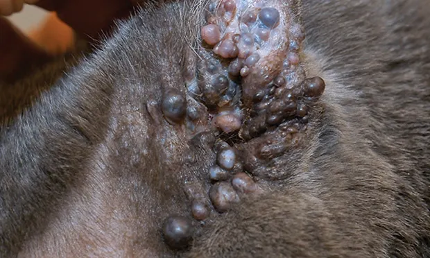
Clinically, lesions are striking and consist of multiple discrete to coalescing blue-gray or purple papules, vesicles and/or nodules (Figures 1 & 2). On rupture of cysts, a yellow-brown to black viscous fluid is easily expressed (Figure 3). Because of the unique presentation, diagnosis is typically straightforward if the clinician is aware of the disease; however, early or small lesions may be mistaken for melanocytic or vascular tumors. Diagnosis is confirmed via histopathology, which reveals clusters of cystic glands.

FIGURE 1
Early ceruminous cystomatosis lesions demonstrating classic punctate blue papules on the concave pinna.
Lesions tend to be cosmetically disturbing for owners, but signs are not usually present if lesions remain small. Ceruminous cystomatosis becomes a problem when lesions enlarge and occlude the canal, which disrupts normal self-cleaning. Disruption of normal otic physiology creates a favorable microenvironment for microbial proliferation which results in secondary otitis externa.
Treatment of ceruminous cystomatosis is typically not warranted unless cysts are enlarging and likely to occlude the ear canal or their presence is resulting in recurrent otitis externa. Ablation of cysts via a carbon dioxide laser is the preferred treatment method; however, surgical excision, cryotherapy, and chemical cautery have been proposed as alternate treatments.
In the experienced practitioner’s hand, laser ablation affords fine control of the treated area, minimizing collateral tissue damage and scar formation, which results in minimal deformation of the pinna.
With appropriate therapy, long-term resolution is quite good, but more than one procedure may be required, depending on the extent and severity of cysts at presentation.