Fecal Sample Analysis
Max, a 6-year-old neutered male Jack Russell terrier, developed malodorous diarrhea after a weekend visit to a sheep farm.
History. Max had no prior medical history.
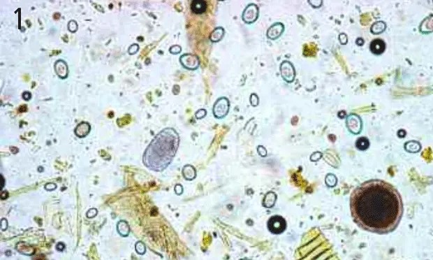
Physical Examination. Max's physical examination was unremarkable. His attitude and behavior were normal. The owner brought along a sample of foul-smelling liquid feces. A centrifugal fecal flotation examination was performed, and the technician reported the presence of coccidia, roundworms, and hookworms. The veterinarian reviewed the slide (Figure 1) and decided to send the remaining fecal sample to a reference laboratory for a second opinion.
Diagnosis: Fecal Artifacts
Fecal material often contains artifacts designated as pseudoparasites and spurious parasites. Pseudoparasites are nonparasitic material that looks parasitic. Common pseudoparasites include plant material (Figures 3 and 4), pollen grains, and free-living arthropods (Figure 5). Spurious parasites are true parasites of one species found in the feces of another species because of coprophagy or predation. In this case, both a pseudoparasite and spurious parasites were present, most likely from the ingestion of sheep manure. Ruminant coccidia oocysts and nematode parasite eggs in the manure are passed undamaged through the gastrointestinal tract. Technical staff trained to recognize only small animal parasites will understandably identify these eggs and oocysts as canine parasites. The structure that looks like a Toxocara egg is a pseudoparasite.
Figure 3. Plant hairs—long, cylindrically shaped structures—are very common fecal artifacts. Although they resemble larvae, they lack organized internal structures. A small, oval-shaped Eimeria oocyst is present in the corner (original magnification ¥400)

Figure 4. There are 1 large and 2 small, dark, round structures, consistent with air bubbles; a transparent glass chip just off center; and a yellowish fragment of plant material (original magnification ¥400).

Figure 5. Grain mites can be present in large numbers in stored dry dog or cat food and can be found on fecal examination (original magnification ´100).

When one is faced with potential artifacts in fecal samples, helpful questions to ask the owner include the following:
Does the pet hunt? Parasites from prey animals can pass through the digestive tract and appear in the pet's feces.
Does a dog have access to fecal material of other animals, including cats, farm animals, and wildlife?
Is the fecal sample fresh? Samples that are not collected immediately after defecation may contain free-living nematodes and mites, as well as maggots.
Size is also an important criterion for evaluating possible fecal artifacts. In this case, the object that looks like a roundworm egg is too big to be Toxocara. The easiest method of measurement is through use of an inexpensive (less than $100) eyepiece reticle. A reticle is a small glass disc with a scale (usually 50 divisions) that fits into a microscope eyepiece. Approximate sizes can be established by using common parasite eggs as a reference. For example, from a standard text you know that eggs of the canine whipworm, Trichuris vulpis, are 70 to 90 µm long. If whipworm eggs measure 7 reticle divisions long on your 10× objective, then each reticle division equals 10 to 13 µm. This calculation can be performed for each objective lens.
In cases for which identification of structures is uncertain, the owner should be instructed to prevent the pet from ingesting nonfood materials and to have a second sample examined after a few days.
ASK YOURSELF ...
What parasites do you see in this fecal flotation (Figure 1)?
Given Max's history, is it possible that the findings are not canine parasites?

DID YOU ANSWER ...
This flotation (Figure 1) appears to contain hookworm (black arrow) and roundworm (arrowhead) eggs and coccidia oocysts (red arrow).
Yes, and indeed these structures are not canine parasites. The structure that appears to be a Toxocara egg is a pseudoparasite, which is indicated by its large size (approximately 120 µm). Also, the outer layer is uneven, with several discontinuities. The egg that resembles the hookworm egg comes from a related family but is an ovine nematode egg. It is larger than the common Ancylostoma species egg, and the presence of other ruminant parasites makes it unlikely that it is an egg of the less common canine hookworm, Uncinaria. On higher-power magnification (Figure 2), the coccidia oocysts can be identified as Eimeria species, not canine Isospora species, by the more oval shape and the presence of the micropyle cap (arrow). Many, although not all, Eimeria species have this cap.