FAST in Emergent Patients
Adesola Odunayo, DVM, MS, DACVECC, University of Florida
Timely assessment of emergent and critically ill patients is necessary to avoid treatment delays, which can increase morbidity and mortality. Focused assessment with sonography for trauma (FAST) has been used as a first-line screening technique for emergencies and as a bedside monitoring tool.
Abdominal FAST consists of 4 standard views—subxiphoid, left flank, midline bladder, and right flank—with the patient in (ideally, right) lateral recumbency. The probe is moved a few inches in several directions and fanned at 45-degree angles until target organs are visualized; approximately 3 to 6 minutes are required to complete all 4 views.
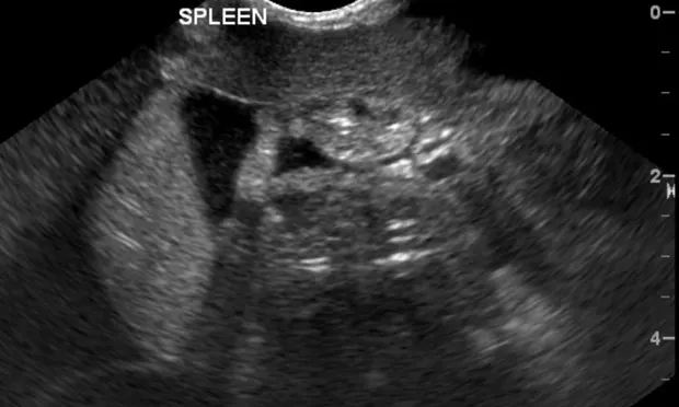
The subxiphoid view aids in assessment of the hepatodiaphragmatic interface, gallbladder, pericardial sac, and pleural space. The left flank view aids in evaluation of the splenorenal interface and areas between the spleen and body wall. The midline bladder view aids in assessment of the bladder apex, and the right flank view aids in examination of the hepatorenal interface and areas between intestinal loops.
Thoracic FAST is performed with patients in left or right lateral recumbency; patients with respiratory distress should be placed in sternal recumbency. The right and left chest tube sites, the right and left pericardial sites, and the abdominal FAST subxiphoid views are evaluated for identification of fluid in the pleural or pericardial space or free air in the pleural space. For the chest tube sites, the probe is held stationary to evaluate for appropriate glide signs, which represent normal lung motion with no chest wall pathology.