Eye Color Change in a Dog
Andrew C. Lewin, BVM&S, DACVO, Vision Vets for Pets, Knoxville, Tennessee
Clinical History & Signalment
Pippin, an 8-year-old spayed Labrador retriever, was presented for a 2-week history of color change in the left eye (OS). The owner reported that Pippin did not exhibit obvious visual impairment, signs of ocular pain, or recent evidence of systemic disease.
Physical Examination
A routine physical examination determined that Pippin was otherwise healthy. A complete ophthalmologic examination was performed and yielded the following results:
Menace response: Normal in both eyes (OU)
Direct and consensual pupillary reflexes: Normal OU
Palpebral reflex: Normal OU
Dazzle reflex: Normal OU
Schirmer tear test: 19 mm/min in the right eye (OD), 21 mm/min OS (OU within normal limits [ie, ≥15 mm/min])
Intraocular pressure: 16 mm Hg OD, 15 mm Hg OS (OU within normal limits [ie, 10-20 mm Hg for rebound or applanation tonometry])
Fluorescein staining: Negative for uptake OU
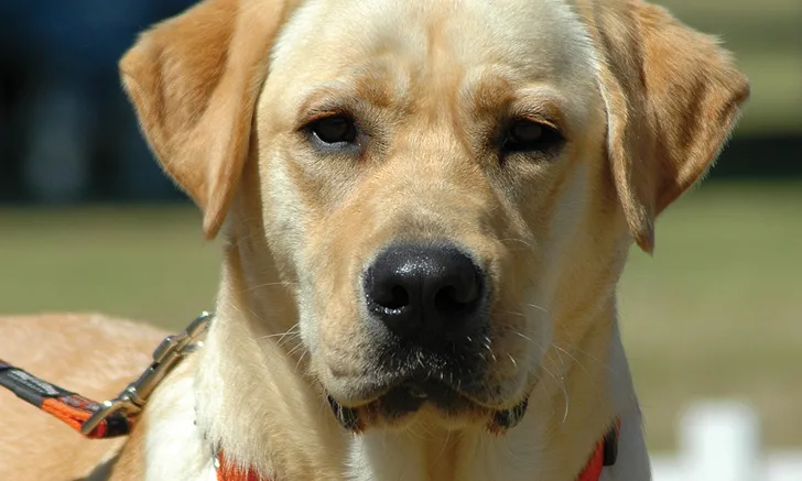
Magnified diffuse beam examination: The anterior segment OD was normal. Two variably pigmented, well-circumscribed, oval to spherical structures were found in the anterior chamber next to the inferior pupillary margin OS (Figure). One structure was smaller in size and immediately adjacent to the larger structure. The remainder of the anterior segment OS was normal.
Magnified slit beam examination: The anterior chamber depth and clarity OD were normal. The abnormal structure OS was easily transilluminated when light was directly applied to the surface, confirming that the structure was a cyst containing clear fluid. No flare was detected OU.
Fundoscopy: Indirect examination using a 28-diopter handheld lens and headset revealed the fundus was normal OU. The abnormal structure in the anterior chamber OS partially obstructed the view.

OS image of the patient. Photo courtesy of Louisiana State University School of Veterinary Medicine Ophthalmology Service
DIAGNOSIS: Uveal Cysts
Diagnosis
Based on ophthalmologic examination findings, Pippin was diagnosed with uveal cysts OS. This is a common diagnosis in dogs and is often a benign and incidental finding.1 The ciliary body is the anterior continuation of the choroid and is connected to the iris. The ciliary processes of the ciliary body are covered in a double layer of epithelium. Uveal cysts are presumed to be of neuroectodermal origin because they typically arise from either the posterior pigmented epithelium of the iris or the inner ciliary body epithelium.2,3 Cysts may be present at the pupillary margin (as in Pippin’s case), posterior iris face, or pars plicata of the ciliary body or be free-floating in the anterior chamber.1 Cysts may change in size over time. A key component to the diagnosis of uveal cysts is transillumination of the cysts. If this is not feasible, ultrasonography with sufficient resolution (ie, ≥50 MHz)4 should be used to differentiate a cyst from a melanocytic neoplasm.1
Treatment & Long-Term Management
In cases similar to Pippin’s in which no visual impairment or concurrent ocular disease is reported, benign neglect is the recommended treatment (see Treatment at a Glance). The owner was asked to bring Pippin back for a recheck eye examination in 6 months, or sooner if any blepharospasm or change in appearance developed.
Treatment has reportedly included either removal of uveal cysts with a needle or deflation with a diode laser in some cases in which cysts interfered with vision.1,5 If treatment is considered for any uveal cyst, referral to a veterinary ophthalmologist is strongly recommended.
TREATMENT AT A GLANCE
In most cases, uveal cysts do not require treatment.
In certain breeds (eg, golden retrievers, Great Danes, American bulldogs), inflammation and secondary glaucoma may accompany uveal cysts. Treatment should be determined on a case-by-case basis; referral to a veterinary ophthalmologist is highly recommended.
Discussion
Uveal cysts in some Great Danes and golden retrievers may represent a component of pigmentary uveitis.6,7 This syndrome is characterized by thin-walled uveal cysts with or without concurrent intraocular inflammation and glaucoma. The disease is common in some populations of golden retrievers and is likely inherited in this breed.8,9 A similar syndrome has also been reported in American bulldogs.10 Examination by a veterinary ophthalmologist may help guide prognosis in some cases.9
Prognosis & Outcome
The prognosis of uveal cysts is good in most cases. Pippin returned to the clinic 12 months later with no change from the previous ophthalmologic examination. The owner reported that Pippin’s vision was maintained.
TAKE-HOME MESSAGES
Uveal cysts are common and usually benign and incidental findings in dogs.
Uveal cysts may be free-floating, attached, fluid-filled, or collapsed against the corneal endothelium or anterior lens capsule.
Certain breeds (eg, golden retrievers, Great Danes, American bulldogs) may present a unique form of the disease.
Uveal cysts can be differentiated from melanocytic neoplasms by transillumination; cysts will often, but not always, allow a slit beam of light to pass through.