Excision of Canine Mast Cell Tumors
Kathleen Ham, DVM, MS, DACVS-SA, University of Florida
In the Literature
Karbe GT, Davis E, Runge JJ, Brown DC, Holt DE. Evaluation of scar revision after inadequate primary excision of cutaneous mast cell tumors in 85 dogs (2000-2013). Vet Surg. 2021;50(4):807-815.
The Research …
Mast cell tumors (MCTs) have variable biological behavior that is difficult to predict and can lead to failed first surgical attempts. Wide surgical excision to achieve a local cure is ideal. An incomplete margin refers to histopathologic findings of neoplastic cells at the cut tissue edge; a narrow margin has no neoplastic cells at the cut edge but a short distance to neoplastic cells. Scar revision surgery with wide surgical excision is recommended when a pathology report indicates incomplete or narrow margins.
This study retrospectively evaluated 85 dogs that underwent scar revision surgery after incomplete MCT excision. Histopathologic assessment of 86 resected scars revealed only 27% had neoplastic cells in the scar, despite incomplete or narrow margins after the first surgery; 8% of resected scars with residual MCT had incomplete or narrow margins.
Follow-up data were available for 68 dogs; 10 had disease progression, and 3 of those developed local recurrence. Margin status and presence of neoplastic cells in the resected scar were not significantly associated with recurrence or progression.
Additional local therapy with surgery or radiation is recommended for incompletely resected MCTs based on a study that demonstrated improved survival times.1 Although none of the patients with incomplete scar revision margins in the current study had recurrence or disease progression, additional local therapy is likely still warranted.
In this study, grade III MCTs were more likely to develop metastasis and disease progression. Specifically, 56% of dogs with grade III MCTs experienced recurrence and/or progression regardless of margin status. Median time to recurrence in 10 patients was 207 days (range, 64-1,583 days); 5 of these dogs were euthanized for reasons related to MCT disease.
… The Takeaways
Key pearls to put into practice:
Dogs with high-grade MCTs are more likely to have disease progression regardless of margin status. Adjunctive treatment and active surveillance should be recommended.
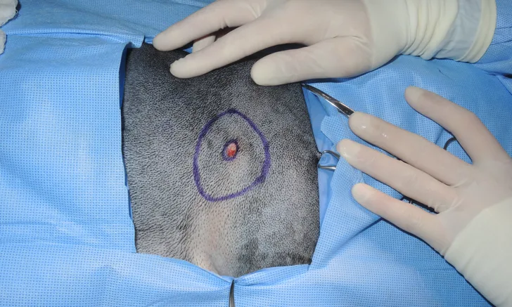
Clinical features of MCTs should be carefully assessed preoperatively to better understand biological behavior. Suspected high-grade MCTs should be excised with wide margins (ie, 3-cm lateral, 1-fascial plane deep; Figure). Knowledge of tumor grade can guide surgical planning and adjunctive treatment and help set pet owner expectations.
All MCTs should be biopsied to assess grade and completeness of excision. Scar revision surgery should be considered if margins are incomplete or narrow.
You are reading 2-Minute Takeaways, a research summary resource presented by Clinician’s Brief. Clinician’s Brief does not conduct primary research.