Evaluation of Chronic Nasal Disease in Dogs: Rhinoscopy in Context
Alasdair Hotston Moore, MA, VetMB, CertSAC, CertVR, CertSAS, MRCVS, University of Bristol, United Kingdom
For the examination of the nose, rigid endoscopy has been shown to be superior to the use of otoscopy cones or flexible endoscopy. However, rhinoscopy by whatever means is rarely appropriate as a sole procedure. Clinicians should recognize that not all regions of the nose, such as the frontal sinuses and maxillary recess, are accessible using this method. In addition, in smaller dogs and cats the nasopharynx is not accessible through the nose, but can be examined by retrograde approach through the mouth.
Other ancillary methods of investigation include:
Radiography (intraoral dorsoventral view, skyline view for frontal sinus; lateral views are rarely helpful)
Magnetic resonance imaging (MRI)/computed tomography (CT) (excellent if available)
Retrograde endoscopy (fiber-optic examination of the nasopharynx)
Serology for aspergillosis
Biopsy of nasal masses
Forced flushing for cytology/histology
Step-by-Step: Nasal Rhinoscopy
What You Will Need
Forward viewing scope (0-10 degrees): 4 mm (medium to large dogs) or < 2.7mm (small dogs and cats) (Figure A)
Light guide and light source• Biopsy (grab/pinch) forceps (Figure B)
Grasping (crocodile) forceps
Optional: Sheath (arthroscopic or urethrocystoscopic), video endoscopy equipment (Figure C)


Step 1
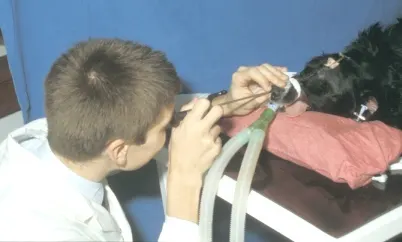
General anesthesia is essential—the procedure is very stimulating and patient movement will cause hemorrhage or more serious damage. Once an endotracheal tube has been placed, position the animal in sternal recumbency.
If irrigation is to be used, pack the pharynx, place the nose in a slightly downward position, and perform irrigation over a bucket or on a gridded tabletop. Using an unsheathed scope reduces the diameter of the scope and may improve access to certain areas of the nose; however, it prevents flushing/irrigation except by infusion through a separate catheter. If an unsheathed scope is used, care must be taken to avoid bending the scope.
Alternatively, you may use an operating sheath over the scope. A sheathed scope allows irrigation and can improve visibility, but it increases the diameter of the scope. In any case, it may be best to look "dry" before attempting irrigation, which may obscure or confuse some features.

Step 2
Examine the more normal of the two sides (judged on radiography or clinical signs) first. Displace the alar fold dorsally and enter the nasal chamber. Examine the dorsal, middle, and ventral meati in turn. Advance only under visual control to avoid damage or hemorrhage.

Step 3
Biopsies should only be attempted once the examination is complete. However, if a foreign body is seen, it is probably best to remove it before further examination provokes hemorrhage. Forceps may be passed via the operating channel but this limits the size of the forcep jaws. Larger biopsy forceps passed alongside the scope will yield a more reliable sample.
Biopsies should be obtained under visual control; however, for large masses (suspected nasal tumors), blind catheter suction biopsies are very useful.• Use a stiff, plastic 8F canine urethral catheter with the end cut off (Figure 3A). The exposed end should be sharp-edged (the sheath of a spinal needle is an alternative).• Under general anesthesia advance the catheter through the nostril while applying suction. Do not enter further than the medial canthus of the eye.• As the catheter tip enters the mass, tissue will enter the lumen of the catheter. If necessary, the tip can be redirected.• The sample can be flushed from the catheter and submitted in formol saline for histopathology (Figure 3B). Epistaxis often occurs but is usually self-limiting.

FIGURE 3A
Procedure Pearl
General anesthesia is essential-the procedure is very stimulating and patient movement will cause hemorrhage or more serious damage.
Procedure Pearl
If a foreign body is seen, it is probably best to remove it before further examination provokes hemorrhage.
Common Chronic Nasal Diseases
Chronic Rhinitis
Description
Common in cats, may be associated with chronic viral infections
Rare in dogs, often described as chronic hyperplastic rhinitis or mucoid hyperplasia
Investigation
Rhinoscopy unrewarding due to large amounts of discharge of variable nature
Radiography is variable in cats. In dogs, destruction of the turbinates and an overall increase in soft tissue density is an important radiographic feature of nasal neoplasia. However, in cats, turbinate destruction and increased soft tissue density can be caused by either inflammatory or neoplastic conditions.
Biopsy may reveal increased quantity of mucus-secreting glands.
Treatment
Response to medical or surgical treatments can be disappointing

Rhinoscopic view of hyperplastic rhinitis in a dog showing an increase in mucus (arrowhead) within the nasal chambers and no abnormal tissue. The turbinates are normal. In the normal dog, there is only scant mucus present in the nasal chambers.
Foreign Bodies
Description
A common cause of sudden-onset sneezing and nasal discharge in dogs
Investigation
Rhinoscopy can be unreliable but may be aided by pretreatment with antibiotics and steroids to reduce discharge
Radiography is less helpful
Treatment
Some foreign bodies dislodge naturally, others may require removal by vigorous flushing under anesthesia or forceps retrieval using endoscopy. Occasionally, open surgery (rhinotomy) is indicated.

Rhinoscopic view of nasal foreign body (grass seed)
Fungal Rhinitis
Description
Uncommon in cats
A common cause of nasal disease in dogs
Infection with Aspergillus and less commonly Penicillium leads to sneezing, nasal discharge, nasal pain, and nosebleeds (epistaxis)
Investigation
Rhinoscopy may show variable discharge but identification of plaques ("mold on jam" appearance) is diagnostic
Radiography shows turbinate destruction in mid to rostral chambers, with increased lucency
Treatment
Oral antifungal drugs and topical treatments (ie, clotrimazole soaks) are the preferred treatment regimen. Twice daily administration of enilconazole (through indwelling catheters placed in the frontal sinuses) is an effective treatment but requires hospitalization and intensive nursing. It is now rarely used.

Rhinoscopic view of nasal aspergillosis. Note the relatively small fungal plague in the center with overlying mucus (arrow) and overall impression of turbinate destruction.
Neoplasia
Description
Common cause of chronic nasal disease (discharge, obstruction, and epistaxis) in elderly dogs and cats
Investigation
Rhinoscopy may be obscured by discharge, but a pale mass with prominent superficial vessels is typically seen
Radiography shows increased radiodensity with turbinate loss ("ground glass" appearance)
Treatment
Surgery alone is not a helpful treatment. For dogs, radiation therapy with surgery is recommended; chemotherapy is recommended for cats

Rhinoscopic view of nasal carcinoma in a dog