Endoscopy of the Avian Respiratory Tract
Bob Doneley, BVSc, FACVSc (Avian Health), West Toowoomba Veterinary Surgery,Toowoomba, Queensland, Australia
Endoscopy-the examination of the interior of body organs, joints, or cavities through an endoscope-had its beginnings in the early 1900s when lighted telescopes were first used to examine inside a human patient. Since its inception, endoscopic examination has increased significantly in both human and veterinary medicine. New endo scopic techniques and equipment are constantly being developed to improve diagnostic ability. Veterinary endoscopy is relatively new when compared with human endoscopy, but veterinary medicine has a wide range of patients that can benefit from this technology.
Avian Endoscopy
Endoscopes were first used in avian medicine in the 1970s and 1980s to determine the sex of parrots, many of which are sexually monomorphic (ie, there are no distinguishing physical differences between the sexes). Examination of the gonads, located between the lung and kidney, allows rapid, accurate identification of sex and revolutionized the breeding of many species. It was quickly realized that the unique avian anatomy, with its air-filled body cavities, made birds an ideal candidate for endoscopy. Insufflation is not required, reducing costs and technical difficulty. Techniques and instruments have been developed to take advantage of this, and endoscopy is now a widely used diagnostic tool in avian medicine around the world.
Indications
Respiratory distress or disease is not uncommon in avian medicine. The following clinical signs may indicate the presence of respiratory disease:
Open-mouthed breathing, of either acute or chronic onset
Increased respiratory effort, seen as tail-bobbing, increased sternal lift, and wing-drooping
Abnormal respiratory noises or change in voice
Radiologic evidence of respiratory disease, such as visible air sac lines, lesions within the lungs or air sacs, reduced air sac space.
Endoscopy should be considered as a diagnostic tool in these patients in the early stages of the disease rather than waiting for a response to other therapies. Severely compromised patients may need to be stabilized before undergoing endoscopy; such interventions as oxygen therapy, antibiotic or antifungal therapy, air sac catheterization, and fluid support may be required.
Step-by-Step: How to Access the Avian Respiratory Tract for Endoscopy
What You Will Need
Endoscope. Most practitioners use a 2.7-mm rigid endoscope with a 30-degree oblique view. This allows a much wider field of view than the more common 0-degree scopes. 1.9-mm endoscopes are available for smaller patients (< 100 g).
Light cable and light source. There are two types of light sources available, the tungsten-halogen and the xenon. Xenon light sources, while more expensive, provide a more intense, whiter, and cooler light than tungsten-halogen. Not only does this give better illumination while viewing inside the body, it is essential if cameras and videodocumentation are to be used. Videodocumentation is desirable, as it allows better documentation of procedures and is a valuable tool for educating both veterinarians and clients.
Endoscopy camera. The use of an endoscopy camera and monitor provides better magnification of the operating field while being more ergonomic for the operator. A 3-chip digital camera gives a high-quality picture and is lightweight and easy to use while attached to the endoscope.
Operating sheath and instruments. The Taylor Operating Sheath (Karl Storz) provides three 5-French instrument channels to allow the introduction of a variety of 3-French endoscopic instruments, including biopsy and grasping forceps, scissors, and aspirating and injecting needles.
Procedure Pearl
A 30-degree endoscope allows a much wider field of view than the more common 0-degree scopes.


Approaches
The anatomy of the avian respiratory tract allows endoscopic examination through both the trachea and the thoracic and abdominal air sacs. The choana and nasal cavity can also be explored endoscopically in some patients. It is important to note that for anything other than a quick examination of the trachea and syrinx, the patient should have an air sac catheter placed to provide anesthesia and respiratory support during the procedure. The approach to placing this catheter is the same as in abdominal endoscopy and will be described shortly.
Tracheal Approach
Under anesthesia, place the patient in either dorsal or ventral recumbency with the head and neck extended. Introduce the endoscope over the tongue and through the glottis into the trachea (Figure 1). Be sure to keep the trachea extended and straight and that the patient is in a surgical plane of anesthesia to avoid iatrogenic trauma. Also keep in mind that the tracheal cartilage in birds is a complete circle-and therefore the trachea cannot be stretched from the inside-and that the lumen of the trachea narrows as it passes caudally to the syrinx. If the syrinx cannot be visualized, it is sometimes helpful to withdraw the endoscope, reverse the patient's positioning (i.e., dorsal to ventral or vice versa), and then reintroduce the endoscope.


Coelomic Approach
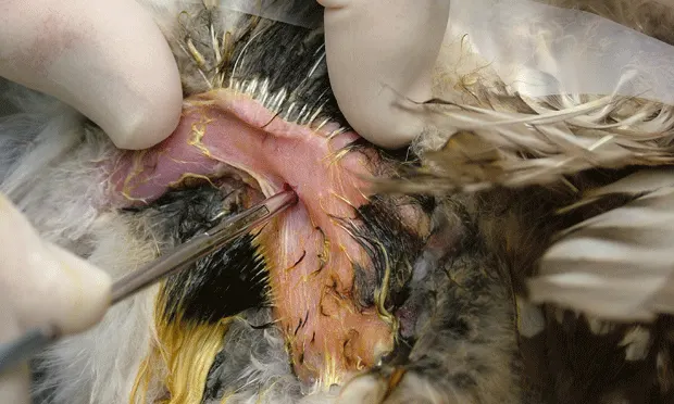
The standard positioning for a coelomic approach is right lateral recumbency with the wings extended and restrained (Figure 2A). Pull the left foot forward and secure it to the neck, and then aseptically prepare the left flank-it is rare to have to remove more than a few feathers.• Identify the following landmarks (Figure 2B): the caudal rib (green line), the flexor crura medialis muscle (red), and the pubic bone (white).• Make a 4-mm vertical incision in the skin (Figure 2B; blue) where the flexor crura medialis muscle crosses the caudal rib (Figures 2C and 2D). Use curved hemostat forceps to reflect the flexor muscle dorsally.• Bluntly dissect the abdominal wall. (Figure 2E) If done correctly, the operator will feel a "pop" as the hemostat forceps enters the caudal thoracic air sac. (If placing an air sac catheter at this stage, a 2- to 3-mm, sterile, noncuffed endotracheal tube can be introduced through the incision into the air sac and secured to the skin.)• Once the coelom has been entered, withdraw the hemostat and introduce the endoscope (Figure 2F).


If entry has been done correctly, the endoscope will now be in the caudal thoracic air sac-this can be confirmed by a clear view of the lung and ostium, with air sac membranes visible on the left and right of the view. The endoscope can be advanced to the lung and, in some patients, can enter the lung through the ostium. The air sac membranes on either side of the caudal thoracic air sac can be penetrated either with endoscopic scissors or by placing the tip of the endoscope against the membrane and twisting sharply. Going to the left allows entry into the cranial thoracic air sac; going to the right allows entry into the abdominal air sac.
Withdraw the endoscope on completion of the procedure. The compromised punctured air sac membranes heal well without any requirement for surgical closure. The muscle and skin incisions can be closed if the operator feels it is advisable; however, they are often left to heal by secondary intention, which occurs rapidly.
Choanal Approach
Introducing the endoscope through choanal slit allows visualization of the left and right nasal cavities and nasal turbinates. Flush saline through the external nares to provide a better examination and to flush out any foreign bodies, or at least out into the field of view. The patient must be intubated before this is performed. If mucus is occluding the endoscope, suction may be used to clear the cavity of excess fluid.
Endoscopic Procedures
Once the area of interest has been visualized, a variety of diagnostic and surgical techniques can be performed. Biopsy samples can be collected for culture and/or histopathologic evaluation, foreign bodies and granulomas may be removed, and occasionally small lesions can be ablated by radiosurgery.
Regardless of the procedure, endoscopy provides a minimally invasive approach with low morbidity and rapid recovery. Patient discomfort is minimal even though extensive diagnostic procedures may have been done.
Conclusion
Endoscopy of the avian respiratory system is a comparatively simple but richly rewarding procedure. It allows direct and magnified visualization of previously inaccessible areas. Biopsies, surgical procedures, and foreign body retrieval are feasible, allowing for much-improved patient care.