Emergency Fracture Management at a Glance
Patients presenting with fractures should receive a complete physical examination, and radiographs should be taken to determine the extent of damage. When the underlying cause is traumatic or suspected to be neoplastic, thoracic and abdominal radiographs and full blood work are recommended. Blood work is also required for fractures requiring surgical stabilization. A CBC, chemistry panel, urinalysis, and/or coagulation profile may be required to safely anesthetize the patient. Taking these steps allows veterinary team members and clients to make the best treatment decisions.
PATIENT TRANSPORTATION
The client should be advised on how to safely transport his or her pet to the practice:
Small pets should be contained in carriers.

The patient can be lifted using a blanket stretcher; a sling can assist in ambulation.
The client may place a towel over the patient’s head to minimize the risk of biting.
Open wounds should be covered with clean towels to minimize contamination.
The client should allow practice team members to transport the patient into the practice, which can be done safely and quickly with a wheeled cart (Figure 3).
Figure 3. A wheeled cart can help safely transport fracture patients. Also, a muzzle allows for safe assessment of the patient.
PRESENTATION
Ask the following questions on presentation:
When and how did the fracture(s) occur?
Are there any concurrent disease processes?
Is the patient on any medications?
EXAMINATION
Assess the patient’s airway, breathing, and circulation, and examine for evidence of hemorrhage.
Check vitals and complete initial diagnostics (TPR, weight, blood pressure, PCV/TS/BG, ECG, SpO2; see Acronyms).
Complete physical, orthopedic, and neurologic examinations.
Obtain radiographs of the affected area.
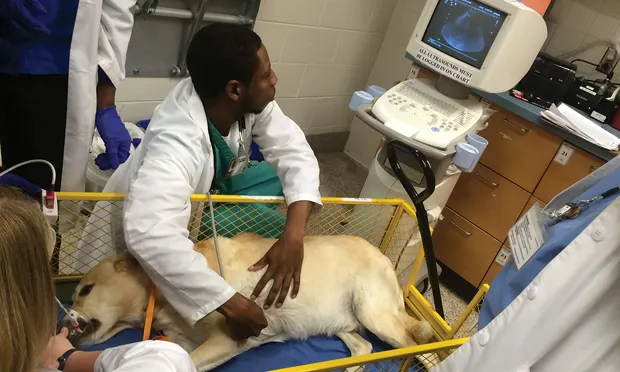
Consider obtaining radiographs of the abdomen, thorax, and spine. Abdominal and thoracic ultrasound may be necessary to detect hemorrhage (Figure 4).

If open wounds and/or fractures are present, obtain a deep tissue sample for culture, then administer broad-spectrum antibiotics.
Evaluate the CBC, chemistry, and coagulation panel (if available) to determine the patient's overall health status.
Figure 4. Abdominal and thoracic ultrasound are sometimes necessary to detect hemorrhage or other serious pathology following trauma.
TREATMENT
Stabilize the patient as needed (eg, place IV catheter, administer oxygen and fluids).
Administer pain medications and sedatives as needed.
Consider using opioids in conjunction with NSAIDS for multimodal pain management. NSAIDS should be avoided in patients who are hypotensive, hypovolemic, or in shock, or whose blood work shows evidence of renal or hepatic disease. NSAIDS should also be avoided in patients who are already on drugs that could cause adverse reactions.
Sedation will facilitate further diagnostics/imaging.
Once analgesics are given, apply sterile lubricant and clip the wounds. Consider collecting samples for culture before cleaning. Flush the wounds with a copious amount of 0.9% buffered sterile saline, which will decrease bacteria by 90%; clean with an antimicrobial. Cover with nonadherent, sterile dressing.6
Discuss assessment, treatment options (eg, external or internal fixation, cage rest, external coaptation, amputation, referral surgery), prognosis, and finances with the client.
If repair is delayed, temporarily immobilize lower limb fractures with a bandage or splint to improve patient comfort and decrease further soft tissue trauma.
TEAM SAFETY
Muzzle patients to allow for safe assessment of wounds.
Wear examination gloves to minimize contamination of open wounds and to protect team members.
| BG = Blood GlucoseECG = ElectocardiogramPCV = Packed Cell VolumeSpO2 = Oxygen SaturationTS = Total SolidsTPR = Temperature, Pulse, Respiration |