Effusion Testing & Classification
Jonathan Bagwell, DVM, DACVP, Dallas Veterinary Specialists, Dallas, Texas
Sample submission, basic classification, further testing for more detailed classification, and refining differential diagnoses are important in point-of-care and laboratory analysis of cavitary effusions. Provided reference intervals typically apply to cats and dogs but can be used as guidelines for other domestic species.
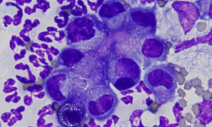
Effusions are classified based on protein content, cellularity, and cytologic criteria. Additional testing (eg, biochemical analysis) can be used for further classification. Primary classifications of effusions are pure transudate, high-protein (ie, modified) transudate, and exudate. More specific classifications include neoplastic effusion, chylous effusion, hemorrhagic effusion, and uroperitoneum.
Pure Transudates
Pure transudates have low protein (<2.5 g/dL) and low cellularity (<1,500 nucleated cells/µL) and are most likely secondary to decreased oncotic pressure associated with reduced serum albumin (often, <1 g/dL).1 Rarely, a pure transudate can be seen in the acute phase of uroperitoneum. In the subacute phase, free urine in the abdomen irritates visceral and parietal surfaces, and subsequent inflammation results in a more exudative effusion.
High-Protein Transudates
High-protein (ie, modified) transudates have moderate to high protein content (2.5-7.5 g/dL) and moderate cellularity (1,000-7,000 nucleated cells/µL).1 This category of effusion is the least specific and most commonly diagnosed in the clinical setting. High-protein transudates are generally caused by increased hydrostatic pressure, increased vascular permeability, or decreased lymphatic drainage. Potential causes include hypertension, hyperthyroidism, lymphatic blockage (eg, associated with neoplasia, granulomas, abscesses), cardiac disease, neoplasia, FIP (typical fluid protein concentration, >4 g/dL), uroperitoneum, visceral organ inflammation, vasculitis, tissue necrosis, lung lobe torsion, and diaphragmatic hernia. Chronic pure transudates can progress to modified transudates due to inflammation associated with the presence of free fluid.
Exudates
Exudates have high protein (>3 g/dL) and high nucleated cell counts (>7,000 nucleated cells/µL)1 and are typically associated with diseases (eg, sepsis, visceral organ inflammation, tissue necrosis, neoplasia, bile peritonitis, uroperitoneum) that cause increased vascular permeability or inflammation. Neutrophils typically predominate and often become degenerate with sepsis; however, lack of degenerate change does not rule out infectious causes. Culture is warranted for most exudates.
*Byline reflects the author’s affiliation at the time of writing. The author’s current affiliation is with IDEXX Laboratories.