Distichiasis
Caryn Plummer, DVM, DACVO, University of Florida
Distichiasis is the abnormal presence of cilia (eg, eyelashes) in the meibomian glands.1
Distichia hair follicles are found in the base of the meibomian gland, where the cilia exit through its orifice; the cilia may contact the cornea and cause irritation. The majority of dogs with distichia do not require any treatment; however, in cases associated with discomfort, therapeutic intervention should be pursued. If the hairs emerge from the conjunctival surface as ectopic cilia, therapeutic removal is usually necessary. Distichiasis is thought to be inherited, but the mechanism of transmission is unknown.2
Any individual patient may present with distichiasis; however, certain breeds are more prone to the condition: American and English cocker spaniels, Welsh springer spaniels, Cavalier King Charles spaniels, flat-coated retrievers, English bulldogs, shih-tzus, poodles, dachshunds, and Parson Russell terriers.
Clinical Signs
Many cases are noted incidentally when an ophthalmic examination is conducted. Others are diagnosed when the patient presents with mild-to-moderate ocular discomfort (eg, persistent or intermittent squinting, increased blinking frequency) and epiphora with or without tear staining on the face.
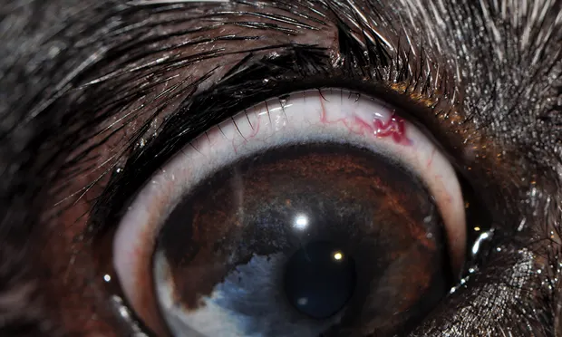
Diagnosis
Distichia are usually noted when a thorough examination of the eyelid margins and ocular surface is performed. Some hairs are large or numerous enough to be noticed by the naked eye; others require magnification to be observed (Figure 1, Figures 2 and 3, below).

FIGURE 1
Multiple distichiae of the upper and lower eyelid of a dog.
Treatment
If hairs are soft and nonirritating, benign neglect is preferred; treatment, if not performed with care, may result in irreversible damage to eyelid margins and meibomian glands, which could result in permanent eyelid deformity or qualitative tear film deficiency.
If the cilia cause discomfort or exacerbate corneal erosions or ulcerations, surgical removal or destruction of the distichia hair follicle is warranted. The simplest treatment for distichia is manual epilation at regular intervals (ie, 4–6 weeks) with smooth-tipped cilia forceps; however, patients with clinical signs as a result of the offending hairs are recommended to undergo surgery for a more permanent solution. To this end, a variety of surgical interventions are available. The most common and, in general, least risky option is cryoablation of the hair-producing follicle (Figure 4, above).3 Cryotherapy is usually performed with either a nitrous oxide or liquid nitrogen probe applied to the palpebral conjunctiva over the affected meibomian glands until an iceball forms that advances across the meibomian gland openings (Figure 5, above). The tissue is permitted to thaw completely and treatment is repeated, followed by manual epilation of any visible distichia. The amount of time necessary to freeze the follicle varies with the cryogen used and thickness of the patient’s eyelid.
Pretreatment with a systemic NSAID may minimize postoperative swelling and discomfort. Postoperative treatment should include systemic NSAIDs and topical corticosteroids. Eyelid and conjunctival swelling after the procedure is common; occasionally, eyelid depigmentation may occur. Other surgical options include electroepilation and surgical excision.
Distichia are similar to other hairs or to cilia in that the hair follicle undergoes a cycle of growth and shedding. Although hairs may not be present in other glands at the time of treatment, they could appear later and require additional treatment.