Digital Dental Radiology
Dental radiology is quickly becoming the standard of care in veterinary dentistry. This is due to the fact that it is crucial for proper patient care and because of a significant increase in client expectations. Providing dental radiography as a routine service can create significant income for a veterinary practice.
Indications for Digital Dental RadiographyIndications for dental radiographs are numerous; a brief list is presented here.
Periodontal DiseaseDental radiographs:
Help elucidate periodontal pockets that may be missed during probing.1
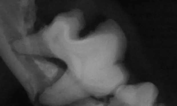
Are absolutely critical in cases of periodontal disease affecting the mandibular teeth of cats and small/toy breed dogs to avoid an iatrogenic fracture during extraction (Figure 1).
May be recommended when several areas of the mouth demonstrate periodontal disease in order to discern other areas affected by subclinical periodontal changes.

Figure 1: Bisecting angle intraoral dental radiograph of the mandibular right arcade of a Yorkshire terrier with significant periodontal disease. Note the significant alveolar bone loss (only 0.3 mm of remaining bone) and secondary weakening of the mandible in the area of the mesial root of the first molar (red arrow). In addition, note the complete bone loss around the mandibular third premolar (white arrow), which is being held in the arcade by a calculus “bridge” to the fourth premolar. In this case, there is significant risk of iatrogenic fracture if extraction is not performed with utmost care.
Feline Tooth ResorptionDental radiographs are essential for diagnostic and therapeutic purposes regarding tooth resorption.<sup2–7sup> There are 3 recognized types of tooth resorptions (see Radiographic Features of Tooth Resorption).
Type 2 lesions demonstrate significant replacement resorption which makes extraction very difficult. In these cases, endodontic infection does not occur, which has resulted in the accepted therapy of crown amputation, but ONLY if there is significant ankylosis and root resorption.
Crown amputation is not an appropriate therapy for type 2 lesions if there is evidence of periapical lucency, periodontal attachment loss of the affected tooth, or generalized stomatitis.
If tooth roots have not resorbed, a complete extraction must be performed.
Endodontic Disease

The most common instance of camouflaged endodontic disease is an uncomplicated crown fracture, where the dentin but not the pulp is exposed (Figure 2).1,2 These teeth are usually vital, but in some cases the endodontic system may become infected through the dentinal tubules.* These painful infections cannot be diagnosed without dental radiographs.

F__igures 2A & 2B: Dental picture of the maxillary left fourth premolar (#208) in a dog with an uncomplicated crown fracture (A). The dental radiograph (B) reveals periapical rarefraction and widened canals indicative of nonvitality and endodontic infection.
Missing TeethIt is very common for teeth to be absent in the dental arcades of veterinary patients.8,9 In some cases the tooth is truly missing, but in others, the tooth/root is present and may be pathologic (eg, retained and infected roots, development of dentigerous cysts).
CR = semidirect; DR = direct; PSP = photo-stimulative phosphor
Other IndicationsAdditional indications for dental radiographs include:
Extraction of deciduous teeth in order to determine root structure8
Jaw fractures1,10
Oral growths/masses
Investigation of possible sources of oral pain
Pre- and postextraction
Several studies have demonstrated the benefit of full-mouth radiographs.11
The Physics & Technology of Digital Radiology SystemsThe 2 types of digital systems are semidirect (CR) and direct (DR).12,13
CR systems employ a photo-stimulative phosphor (PSP) plate covered with phosphor crystals, which temporarily store the x-ray photon energy. Following exposure, the film is removed from the patient’s mouth and scanned. The scanner then transfers the information to the computer for processing.
DR systems employ solid state sensors, which are linked via a USB cable or Bluetooth to the computer, resulting in the image being available in seconds.
Computers have the ability to manipulate images in order to change their appearance. This manipulation is performed by subjecting the information to a mathematical algorithm. Algorithms can:
Improve contours or contrast
Adjust an image that is too light or dark.
These algorithms are responsible for many of the differences in the appearance of images between manufacturers. In many cases, the raw image is very similar but the algorithm gives it a slightly different appearance.
There are no published studies that demonstrate a significant difference between the digital images produced by the various sensors. Most studies report that while solid state sensor systems have a higher resolution, the overall image quality is equal. The prospective purchaser should evaluate all available systems before making their decision; one website that presents a comparison is vetdentalrad.com.
Advantages of Digital Dental Radiography

SpeedThe major advantage of digital dental radiography is speed. With the DR systems, the image is available in seconds. Additionally, since the image is produced while the sensor and tube head are still in position, adjustments to the angle can be made without starting over (Figure 3). In contrast, the need to scan PSP plates makes CR systems much slower than DR systems; however, PSP plates are still faster to develop than standard film.
Figure 3: Photograph of patient under general anesthesia. Note that the radiographic image appears while the sensor and tube head are still positioned. This allows minor technique adjustments to be made quickly when necessary.

Interpretation of ImagesWith both types of digital systems, the large size of the image projected on the computer screen, coupled with the ability to manipulate the image, allows for easier interpretation (Figure 4). The large computer screen image is also very helpful during discussions with clients, and offers the ability to “mark” the images to further facilitate client educational communication (Figure 5).
Figure 4: Intraoral dental radiographs of the right and left mandible of a cat. The patient has squamous cell carcinoma on the left, which has caused significant bony resorption (red circle). The right side is shown on the same screen for comparison (with an arrow pointing to the intact mandibular cortex). In addition, there is advanced type 2 tooth resorption on the mandibular right third premolar (#407) (shown by another arrow). The ability to mark the images, along with side by side comparisons of the contralateral “normal,” is very valuable when educating clients.

Figure 5. The large format of digital dental radiography images and ability to mark the images greatly improve client communication. In this radiographic image, the mandibular right third premolar (#407) is nonvital as evidenced by the wide root canals. (This tooth is marked with a red arrow.)
Telemedicine & Other AdvantagesThese images can be quickly uploaded to telemedicine sites for specialist review. Additional benefits include:
Permanence of digital images (if backed up on computer systems)
Avoiding use of toxic chemicals
Less storage space required
General ease for staff to locate.
DR systems have an added advantage of creating far less radiation exposure compared with standard film. CR systems can potentially have less radiation as well, but their radiation range is much more variable.
Disadvantages of Digital Dental RadiographyDetail of ImageThe main criticism of digital dental radiography has always been that they have less detail than film. However, improvements in computer monitors, imaging capture hardware, and image manipulation software have improved the quality of digital dental images to the point where many current products on the market have at least the same detail as standard film.
Sensor Plates

The biggest drawback of DR systems is the lack of a size-4 sensor plate. This typically only presents a problem in large-breed dogs in which the canine and carnassial teeth cannot be completely imaged on a size-2 sensor (Figure 6). The larger size-4 plate available with the PSP system decreases the amount of exposures needed to take full-mouth radiographs.
Figure 6: Bisecting angle intraoral image of the mandibular canines in a large-breed dog obtained with a size-2 sensor. Note that while the entire sensor was utilized, only the roots were imaged (and not completely).
Distribution, Storage, & ManagementThe requirements for image distribution, storage, and management are growing in the veterinary practice. This has resulted in the development of picture archiving and communication systems (PACS) to facilitate these procedures. An open standard called DICOM (digital imaging communications in medicine) is the de facto standard for imaging in human medicine and is ubiquitous with veterinary whole-body radiography, ultrasound, MRI, and CT, and is nearly ubiquitous with veterinary practice information management software (PIMS). It makes communication and locating images for both the practice and telemedicine much easier.
Unfortunately, to date, true DICOM compliance is not readily available with digital dental systems; most of them use proprietary bridges. However, most manufacturers are making significant advances in this area; ask your representative about their system’s DICOM compatibility prior to purchasing. While this may not be a deciding factor, it should be taken into consideration.
Economic ImpactThe biggest concern when converting to digital dental radiology is the initial set-up cost. However, there are significant cost savings in film and development. Furthermore, the speed of diagnostic image acquisition and a greater compliance of veterinary staff to use this equipment more than compensate for these initial costs. A busy general practice should easily make up the difference between film and a digital system within one year, at which point the digital system becomes less expensive.
ConclusionDigital dental radiology is the future of veterinary dentistry. It is more efficient, requires less radiation, utilizes no toxic chemicals, allows for easy telemedicine consults and better communication with clients, costs less than standard radiographs over time, and offers permanent image storage.
CT = computed tomography; DICOM = digital imaging communications in medicine; DR = direct; MRI = magnetic resonance imaging; PACS = picture archiving and communication systems; PIMS = practice information management software
DIGITAL DENTAL RADIOLOGY • Brook A. Niemiec & Matthew Wright
References
1. Case based dental radiology. Niemiec BA. Top Comp Anim Med 24:4-19, 2008.
Pathologies of the dental hard tissue. Dupont GA. In Niemiec BA (ed): Small Animal Dental and Oral Maxillofacial Diseases—London: Manson, 2010, pp 127-157.
Domestic feline oral and dental disease. Wiggs RB, Lobprise HB. Veterinary Dentistry, Principals and Practice—Philadelphia: Lippincott Raven, 1997, pp 487-496.4. Diagnostic value of full-mouth radiography in cats. Verstraete FJ, Kass PH, Terpak CH. Am J Vet Res 59:692-695, 1998.5. Comparison of periodontitis and root replacement in cat teeth with resorptive lesions. DuPont GA, Debowes LJ. J Vet Dent 19:71-76, 2002.6. Prevalence of odontoclastic resorption lesions and periapical radiographic lucencies in cats: 265 cases (1995-1998). Lommer MJ, Verstraete FJ. JAVMA 2000 217:1866-1869, 2000.
Crown amputation with intentional root retention for dental resorptive lesions in cats. DuPont GA. J Vet Dent 19:107-110, 2002.
Pathology in the pediatric patient. Niemiec BA. In Niemiec BA (ed): Small Animal Dental and Oral Maxillofacial Diseases—London, Manson, 2010, pp 89-126.9. Clinical, radiographic, and histological characteristics of secondary retention of permanent molars. Raghoebar GM, Boering G, Vissink A. J Dent 19:164-170, 1991.10. Atlas of Canine and Feline Dental Radiography. Mulligan T, Aller S, Williams C—Trenton, New Jersey: Veterinary Learning Systems, 1998, pp 176-183.11. How to obtain and interpret periodontal radiographs in dogs. Tsugawa AJ, Verstraete FJ. Clin Tech Small Anim Pract 15:204-210, 2000.12. Filmless imaging, the uses of digital radiography in dental practice. Van Der Stelt PF. J Am Dent Assoc 136:1379-1387, 2005.13. Digital dental radiography. Niemiec BA. J Vet Dent 24:192-197, 2007.