Differentiation of Parasites & Pseudoparasites

ArticleLast Updated April 20098 min readPeer Reviewed

Identifying stages of parasites in samples prepared from fecal specimens is important for the proper diagnosis of parasitic infections. The best approach seems to be the memorization of the size and appearance of the expected parasites that might be found in the feces of the sampled host. Also, an ocular micrometer or an image with a scale bar is invaluable for determining an item's size, which is critical for making comparisons with images in books or other references.

A conundrum for even a trained technician is the presence of common nonparasitic items that are regular fecal elements (ie, artifacts). Some artifacts are from ingested food, some are from the air, and some are nonpathogenic inhabitants of the intestinal tract.

The pictures presented are of regularly occurring parasite stages and identified artifacts. Choose from the listed diagnoses; each description corresponds with only 1 picture. (See Full Article for Pictures)

See pages 58-59 for answers.

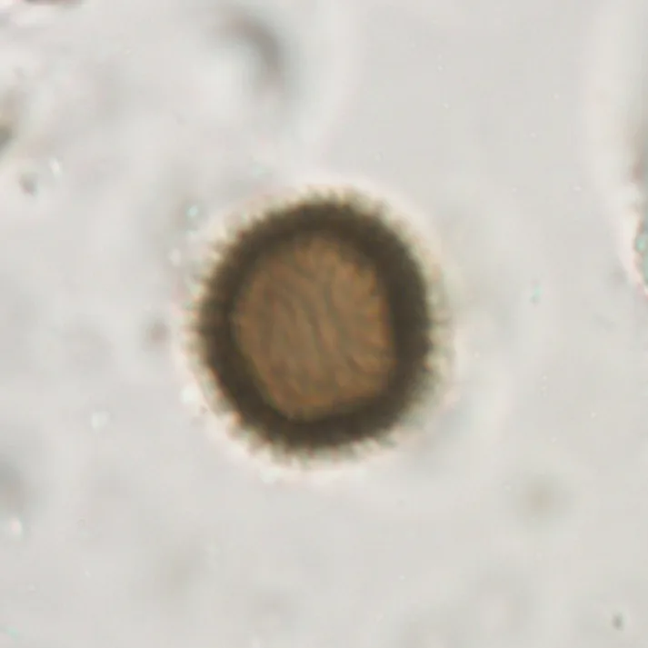
1. Smut fungal spore (possibly a Tilletia species)Smuts grow on the seeds of grasses, most notably grains. The hyphae of the fungi grow in the stem, but ultimately, as the grass flowers, they take over the seed, turning it into a distribution system called a sorus or bunt ball that is full of teliospores. At harvest (or upon ingestion), the sori burst to release the teliospores, which each have a characteristic shape and surface morphology. To identify this specific spore, we had help from Dr. Kathie Hodge (Associate Professor of Mycology, Department of Plant Pathology & Plant-Microbe Biology, Cornell University). This spore was recovered from a ruminant fecal sample; however, we can find similar spores in the feces of almost any host.(bar = 20 mcm)

  1. Fungal conidiumConidia are asexually generated airborne spores of fungi within the phylum Ascomycota. They are often brown and may have one pointed or rounded end; the other end may look as though it has a plug or opening where it was attached to the fungal hypha. Sometimes, a bit of the fungal hypha remains attached. The spores are very common in the air and are regularly ingested or inhaled by animals, coughed up, and swallowed with respiratory mucus. They are often mistaken for the eggs of Trichuris or capillarids because of their brown color and the shape of the poles. Vice versa, if a Trichuris egg is oriented off-axis with one end pointing downward from the coverslip, it may appear to have only one polar plug and look more like a conidium.(bar = 20 mcm)

3. Taeniid eggThe taeniid tapeworm egg is about 30 mcm in diameter and has a shell that appears to be striated. This shell is formed from thousands of 6-sided structures that are narrower at the base than at the surface; you can watch the shell fall apart into its individual units if you add a drop of dilute chlorine bleach to the slide. The eggshell (embryophore) surrounds a hexacanth (bearing 3 pairs of hooklets) larva that is fully formed when passed in the feces. The eggs are infectious when passed. As eggs in the genera Taenia and Echinococcus cannot be readily distinguished from one another, the typical readout is "taeniid tapeworm egg."(bar = 20 mcm)

4. Monocystis lumbrici sporocystsThis is the sporocyst of a gregarine apicomplexan protozoa parasite of the earthworm. These appear in the feces of dogs and other animals that sometimes eat earthworms. The stage passed in the feces is the sporocyst, which contains 8 sporozoites. On some occasions, the sporocysts might be balled together within the large oocyst digested out of the earthworm. These oocysts contain a very large number of sporocysts, whcih in turn have characteristic pointed ends. The typical sporocyst of Monocystis lumbrici is about 15 to 20 mcm long.(bar = 20 mcm)

5. Giardia duodenalis cystThe cysts of this parasite are common in the feces of dogs, cats, and other animals. They can be present in 30% or more of certain populations and are commonly seen on fecal examinations. However, it may be difficult to distinguish them from the sporocysts of Sarcocystis, which are very similar in size and shape. The Giardia cyst will contain 4 nuclei that may or may not be apparent depending on the microscope used and the lighting applied; they are ellipsoid in outline and about 10 to 12 mcm long. The similarly shaped sporocysts of Sarcocystis, if examined carefully, will be seen to contain 4 elongate sporozoites and the sporocyst residuum.(bar = 20 mcm)

6. Hemlock PollenHemlock tree pollen (Tsuga species, within the family Pinaceae) is characterized by being solitary and round. The grains have large blunt and undulating projections covering their surface, often appearing as a "fringe" around a large central depression. They are often greater than 40 mcm in size and appear similar in size, shape, and color to the egg of Toxocara.(bar = 20 mcm)

7. Plant hairMany plants are protected by small hairs. Very typically, hairs have central hollow shafts that may give them the appearance of having internal structures, like the intestine within a nematode larva. These hairs are commonly seen on fecal flotations, and many are similar in size to nematode larvae found in feces. They can be very disconcerting, especially to those who are only recently initiated into the world of fecal examination. We shaved the fuzz from a peach to acquire the hair for this image, but similar hairs abound on different plants eaten by animals.(bar = 100 mcm)

8. Toxocara canis egg (infertile)The eggs of Toxocara have a typical pitted eggshell surrounding a dark fertilized zygote. The darkness of the large central zygote (along with the pitted eggshell) is the main characteristic used to distinguish T canis and T cati eggs from the smooth-shelled eggs of Toxascaris leonina, which contain a light-colored zygote. However, the center of the Toxocara egg will, on occasion, appear lightly colored. Early in life or when males are unavailable, a female Toxocara will produce eggs containing a light-colored unfertilized ovum surrounded by an eggshell that may be less regularly pitted than that of the fertilized egg.(bar = 20 mcm)

9. Saccharomycopsis (Cyniclomyces guttulatus)This yeast, which lives in the intestinal tract of dogs and rabbits, is considered a commensal by most individuals, although some insist it is pathogenic. The yeast colonizes the stomach and intestine of rabbits, and it has been found in the intestines and bile ducts of dogs. The cells multiply by budding, but dividing stages are not typically found in the feces-the stages in the feces are about 20 mcm long and often contain 2 vacuoles. If this organism colonizes the intestine and is not pathogenic, it is called a commensal rather than a parasite or an artifact.(bar = 20 mcm)

10. Sarcocystis sporocystsDogs, cats, and other carnivores pass the sporocysts of Sarcocystis in their feces after having ingested raw meat containing sarcocysts. Gametogony occurs in the intestinal mucosa of the predator, and the stage passed in the feces is a sporulated sporocyst. This stage contains 4 sporozoites and a residual body. The sporocysts are about 10 to 12 mcm long and ellipsoid in outline; therefore, they appear very similar to the cysts of Giardia duodenalis.(bar = 20 mcm)

11. Trichuris vulpis eggThis is the egg of the whipworm that is common in the dog. It is in the single-celled stage when passed in the feces, and often the shell and the contents are dark orange-brown in color. The bipolar plugs are characteristic, and the egg is much larger, about 3 to 4 times the size, of the conidia of various fungi that occur in the feces of animals. Misidentifications may occur when people are not careful about what magnification they are using to examine the egg or are unsure how large the conidium is that they are observing.(bar = 20 mcm)

12. Aelurostrongylus abstrususThis particular larva of the feline lungworm was recovered by using centrifugal sugar flotation. It is damaged less by the flotation process than other nematode larvae, and will often look fairly larva-like soon after flotation. However, in this case, the morphology has been affected by the osmotic pressure exerted by the sugar. Even so, if you have successfully identified this as a larva and are fortunate enough to have a specimen that allows you to convince yourself that you see the kinked tail or dorsal spine, these characters will help distinguish it from other nematode larvae, such as those of hookworms. This larva is about 375 mcm in length. (The insert shows another larva recovered in a sugar float from a cat fecal-the kink in the tail is recognizable, but the dorsal spine is not obvious.)(bar = 100 mcm)