Differentiating Gastric Carcinoma from Chronic Gastritis
Frederic Gaschen, Dr.med.vet., Dr.habil., DACVIM (SAIM), DECVIM-CA (IM), Louisiana State University
In the Literature
Seim-Wikse T, Skancke E, Nødtvedt A, et al. Comparison of body condition score and other minimally invasive biomarkers between dogs with gastric carcinoma and dogs with chronic gastritis. J Am Vet Med Assoc. 2019;254(2):226-235.
The Research …
Gastric carcinoma has a very low incidence in dogs.1 Affected dogs may present a diagnostic challenge, as their history and clinical signs (eg, vomiting, dysrexia, weight loss) are not specific and can overlap with signs of many other, more common chronic diseases affecting the GI tract and other organ systems. A typical diagnostic investigation should include CBC, serum chemistry profile, and urinalysis to screen for diseases outside the GI tract, as well as abdominal imaging (both radiography and, particularly, ultrasonography may be helpful) and gastroscopy with collection and histopathologic analysis of mucosal biopsies.
The authors of this prospective study sought to find noninvasive markers that could help clinicians develop a higher index of suspicion for gastric carcinoma in dogs presented with compatible clinical signs. Three groups (ie, dogs with gastric carcinoma, dogs with chronic gastritis, healthy dogs) were compared. All diagnoses were histologically confirmed using an established grading system. The authors found that dogs with gastric carcinoma tended to be older (median age, 9.5 years; range, 8-15 years) than those with chronic gastritis (median age, 6.5 years; range, 1-12 years) and had a lower BCS (≤4/9 in all dogs with carcinoma for which BCS was recorded). Although there were no clinically relevant differences in CBC and serum chemistry results, easily measurable serum concentrations of C-reactive protein (CRP) were higher and serum folate concentrations were lower in dogs with gastric carcinoma as compared with healthy dogs or dogs with chronic gastritis.
In a recent study, abdominal ultrasonography correctly identified 50% of gastric neoplasms in dogs and cats.2 In this study, abnormalities of the gastric wall and regional lymph nodes were detected in all dogs with gastric carcinoma undergoing ultrasonography. Gastric carcinoma is often localized on the pyloric antrum or along the lesser curvature (Figure). Due to proximity to the abdominal wall, antral wall lesions may be easier to observe than are lesions in the gastric fundus/body, particularly to the less experienced ultrasonographer.
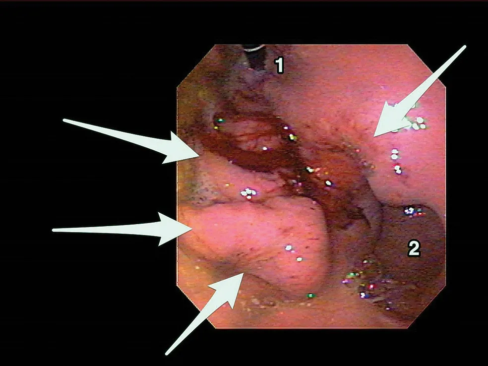
Gastroscopic view of a large, sessile, bleeding mass along the lesser curvature of the gastric fundus in a 12- year-old crossbreed dog with chronic vomiting and weight loss of 1 year’s duration and a worsening condition. The thickened gastric mucosa (arrows), endoscope in the cardia (1), and gastric antrum (2) can be observed. The mass could not be easily detected during ultrasonographic examination due to its location, the uncooperative nature of the dog, and the presence of air in the gastric lumen. Histopathology revealed gastric adenocarcinoma. Photo courtesy of Dr. Ahuja and Dr. Johnston, Louisiana State University
… The Takeaways
Key pearls to put into practice:
Gastric carcinoma is a rare tumor of dogs. Because clinical signs are nonspecific, diagnostic investigation in dogs with compatible signs should include a minimum database (ie, CBC, serum chemistry profile, urinalysis) to screen for more common diseases sharing the same clinical signs.
If gastric carcinoma is further suspected after minimum database, abdominal radiography and ultrasonography may be useful. Final confirmation is made with gastroscopy and histopathology of mucosal biopsies and may be best performed by a specialist.
The clinician should evaluate serum CRP and folate concentrations in patients of older age and/or with a lower BCS when referral is not an option. Although high CRP and low folate levels raise the index of suspicion for gastric carcinoma, confirmatory tests (as previously described) are still required to obtain a final diagnosis.