Differential Diagnosis of Urinary Incontinence
History. Denise, a 12-year-old, spayed female black Labrador retriever, was presented for evaluation of urinary incontinence. The incontinence was most pronounced when she was sleeping or relaxed and lying down. In addition, Denise was having "accidents" in the house associated with pollakiuria, dysuria/stranguria, and hematuria. The incontinence associated with relaxation/sleeping had been present for several months and preceded the house-soiling behavior. In all other respects, the owner felt that Denise was normal.
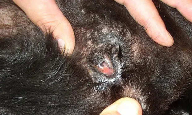
Examination. Denise was slightly overweight and had a moderately "tucked in" vulva; there was some perivulvar inflammation, with evidence of licking and pigmentation change (Figure 1, above). A complete neurologic examination was performed; pudendal nerve sensory and motor functions were thought to be intact, there was no lumbosacral pain, and there were no conscious proprioceptive deficits. Findings on rectal examination of the pelvic urethra and trigone region of the urinary bladder and digital vaginal examinations were normal. Denise's urinary bladder was small and felt normal on abdominal palpation. Resistance to manual bladder expression was thought to be neurologically normal. The rest of the physical examination was unremarkable.
Laboratory Results. A urine sample was obtained by cystocentesis, and Denise was then walked outside to observe her voiding behavior. She postured normally and produced a normal urine stream without apparent dysuria or stranguria. Palpation of the urinary bladder after voiding was normal; the residual urine volume was small and the bladder wall was not thickened. Results of analysis of a urine sample obtained by cystocentesis are presented in the Table. Results of a complete blood count and serum biochemistry profile were within normal limits.
Radiographs. Plain abdominal radiographs were unremarkable.
ASK YOURSELF ...
Which of the following is the best initial assessment of this case?A. Bacterial cystitis causing urge/inflammatory incontinenceB. Bacterial contamination from intestinal tract associated with cyctocentesis samplingC. Primary urethral sphincter incompetence with secondary bacterial cystitisD. Inappropriate urination associated with a behavioral problemE. Inapproriate urination and incontinence associated with polydipsia/polyuria
Correct Answer: C**Primary urethral sphincter incompetence with secondary bacterial cystitis is most likely.**
Assessment. The most commonly encountered forms of urinary incontinence are detrusor instability (urge or inflammatory incontinence) and urethral sphincter mechanism incompetence (USMI). With inflammatory disorders, incontinence occurs because bladder or urethral irritation results in detrusor muscle instability (failure of the bladder to remain relaxed or compliant during the storage phase of micturition). In these cases, the urge to urinate overcomes normal house-training behavior.
On the other hand, USMI frequently occurs in middle-aged to older, large-breed, spayed females. Aging or a relative decrease in estrogen concentrations, which can adversely affect collagenous support structures (and thereby cause decreased sphincter tone), can result in USMI. Other causes include a decrease in the number or responsiveness of α-adrenergic receptors in the smooth muscle of the urethral sphincter. Obesity or vaginal abnormalities may also exacerbate sphincter incompetence or compromise host defense mechanisms and allow secondary urinary tract infection.
History, physical examination, neurologic examination, urinalysis, and response to treatment are used to differentiate USMI from detrusor instability. Historically, USMI is most pronounced when the animal is asleep or relaxed. Owners often report that their pet's bedding is wet when the animal gets up after lying down. In contrast, incontinence with a history of hematuria, pollakiuria, and dysuria or stranguria usually indicates inflammation of the bladder or urethra. Inflammation of the bladder or urethra that results in detrusor instability probably creates a sensation of bladder fullness, which triggers the voiding reflex.
In this case, the patient history and hematuria do not fit well with a behavioral voiding problem, the single organism grown from the urine culture does not support intestinal tract bacterial contamination during the cystocentesis procedure, and the urine specific gravity of 1.031 does not support a diagnosis of polyuria-polydipsia.
Diagnosis. Urethral sphincter incompetence is probably the primary disorder. The patient history suggests that the sphincter incompetence preceded the signs of lower urinary tract inflammation. Decreased internal urethral sphincter tone and the tucked-in vulva probably compromised host defense mechanisms and allowed bacteria to more easily ascend through the urethra, resulting in a secondary bacterial cystitis.
TAKE-HOME MESSAGES
• USMI frequently occurs in middle-aged to older, large breed, spayed females.• USMI can be complicated by ascending urinary tract infections, which can change the clinical presentation. History is often helpful in determining the primary problem.• In addition to the USMI, vulvar abnormalities or perivulvar dermatitis can also predispose the patient to a secondary, complicated urinary tract infection (ie, infection coexisting with compromised host defense mechanisms, in this case abnormal vulvar anatomy, USMI, and perivulvar dermatitis).• USMI frequently responds to α-adrenergic agonists (eg, phenylpropanolamine) or estrogen compounds (eg, DES).• α-Adrenergic agonists and estrogens are synergistic, and combination treatment may produce continence in patients that do not respond to single-agent treatment. In refractory cases, other diagnostic tests, such as urethral pressure profilometry and additional imaging for anatomic abnormalities (ultrasonography or contrast radiography), may be helpful.• Complicated urinary tract infection should be treated on the basis of urine culture and sensitivity for a minimum of 4 weeks or until the host defense mechanism abnormalities are corrected.
TX at a glance
Urethral Sphincter Mechanism Incompetence• α-adrenergic agonist (eg, phenylpropanolamine) or estrogen compounds [eg, DES]• In refractory cases, use both of the above agents
Complicated Urinary Tract Infection• Agent chosen on basis of urine culture and sensitivity test results for minimum of 4 weeks
DIFFERENTIAL DIAGNOSIS OF URINARY INCONTINENCE • Gregory F. Grauer
Suggested ReadingEvaluation of phenylpropanolamine in the treatment of urethral sphincter mechanism incompetence in the bitch. Scott L, Leddy M, Bernay F, et al. J Small Anim Pract 43:493-496, 2002.Symposium: A diagnostic approach to micturition disorders, treating urinary incontinence, and medical treatment of voiding dysfunction in dogs and cats. Lane IF, Fischer JR. Vet Med January:49-74, 2003.The diagnosis of urinary incontinence and abnormal urination in dogs and cats. Silverman S, Long CD. Vet Clin North Am Small Anim Pract 30:427-448, 2000.Urethral sphincter mechanism incompetence. Arnold S, Weber U. In Bonagura JD (ed): Current Veterinary Therapy XIII-Philadelphia: WB Saunders, 2000, pp 896-899.Urinary incontinence. Forrester SD. In Ettinger SJ, Feldman E (eds): Textbook of Veterinary Internal Medicine, 6th ed-St. Louis: Elsevier, 2005, pp 109-111.