Diabetes Mellitus Part 1: Diagnosis
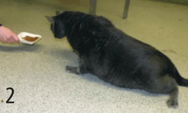
Image above. Some diabetic cats develop a peripheral neuropathy characterized by knuckling of the hindlimbs.
Profile
DefinitionDiabetes mellitus (DM) is a common endocrinopathy characterized by decreased insulin production or decreased insulin function. In both cases decreased insulin action results in hyperglycemia.1
Related Article: Diabetes Mellitus Part 2: Treatment
Systems. Insulin is an anabolic hormone which generally moves amino acids, fatty acids, glucose, and electrolytes into cells, and facilitates cell growth by promoting protein, adipose tissue, and glycogen synthesis. Therefore, decreased insulin production affects all cells in the body.
Lack of insulin action results in muscle and adipose tissue catabolism. Hyperglycemia can lead to glomerular disease (in dogs and cats), cataract formation (dogs), or peripheral neuropathy (cats).2-4
Genetic Implications. The predisposition of some breeds to develop DM indicates that genetic factors exist. Various genes are probably involved in the pathogenesis of canine and feline DM, and the major histocompatibility complex genes may have an important role in the pathogenesis of DM in dogs. Genetic studies have not been reportedin cats.
Incidence/Prevalence.In dogs, 64 of every 10,000 patients admitted to hospitals have DM.5 In cats, the incidence was determined to be 2.45 cases per 1000 cat-years-of-risk during a 6-year study period.6SignalmentBreed Predilection. Samoyeds, Australian terriers, miniature schnauzers, toy and miniature poodles, and pugs as well as Burmese cats are at increased risk for DM. German shepherds, golden retrievers, and American pit bull terriers are at decreased risk.<sup7 sup>
Age. The mean age of onset in dogs is 7 to 9 years; in cats the mean age is 10 years. However, any age dog or cat can develop the disease.
Gender. Female dogs and neutered male cats may be at increased risk for DM.
CausesThe etiology of DM is incompletely understood and is likely multifactorial. In dogs, genetic factors and immune-mediated destruction appear to be important etiologic factors. In cats, amyloid deposition may contribute to development of DM.
Risk FactorsBreed predisposition is an important risk factor. Obesity and carbohydrate-rich diets may increase the risk of DM in cats.
PathophysiologyLack of insulin action results in hyperglycemia. When hyperglycemia exceeds the renal threshold of 180 mg/dL in dogs and 280 mg/dL in cats, glucose reabsorption in the proximal renal tubules is incomplete and glucosuria develops. Glucose acts as an osmotic agent, drawing fluid into the urine and causing polyuria; polydipsia develops to compensate for loss of water in the urine.
In addition to decreased mobilization of glucose into cells, lessened insulin action also leads to impaired mobilization of amino and fatty acids into cells. Polyphagia develops to compensate for this “starvation” of the cells.
SignsHistory. Polyuria and polydipsia, weight loss, and polyphagia are the most common clinical signs observed. Blindness (dogs) and a plantigrade stance (cats) can also develop.
Diabetic cataracts are common in dogs. They are treated surgically; however, it is important to achieve optimal diabetic regulation prior to surgery.Physical Examination. Animals may appear normal or be severely compromised. Findings may include:
Variable body weight: Underweight, normal, or obese
Variable hydration status: Normal or dehydrated
Hepatomegaly
Cataracts (dogs) (Figure 1)
Plantigrade stance (cats) (Figure 2)
Lethargy, weakness, and acetone breath—mainly noted in severely compromised patients with diabetic ketoacidosis
Most diabetic dogs and cats are middle-aged to older patients and may have concurrent disease.2,8 History and physical examination findings may be affected by concurrent diseases such as:Dogs & Cats
Urinary tract infection
Acute pancreatitis
Neoplasia
Mainly Dogs
Hyperadrenocorticism
Hypothyroidism
Mainly Cats
Other infections
Chronic renal failure
Hyperthyroidism
Diagnosis
Definitive DiagnosisAll of the following criteria must be satisfied in order to make a definitive diagnosis:
Appropriate clinical signs and physical examination findings
Persistent hyperglycemia
Glucosuria
Differential DiagnosisDogs & CatsPolyuria/polydipsia:
Renal or liver disease
Hypercalcemia
Hypokalemia
Drug administration/iatrogenic: Glucocorticoids, diuretics, anticonvulsants, fluid overload
Weight loss & polyphagia:
Gastrointestinal parasites
Protein-losing enteropathy or nephropathy
Hyperglycemia:
Drug administration: Glucocorticoids, progesterone, megestrol acetate
Total parenteral nutrition or other dextrose containing IV fluids
Postprandial hyperglycemia9-11
Diestrus
Acute pancreatitis
Factitious measurement
Glucosuria:
Primary renal glucosuria
Mainly DogsPolyuria/polydipsia:
Hyperadrenocorticism or hypoadrenocorticism
Pyometra
Diabetes insipidus
Psychogenic disease
Polycythemia
Weight loss & polyphagia:
Exocrine pancreatic insufficiency
Hyperglycemia:
Hyperadrenocorticism
Pheochromocytoma
Mainly CatsPolyuria/polydipsia:
Hyperthyroidism
Weight loss & polyphagia:
Hyperthyroidism
Hyperglycemia:
Stress
Acromegaly
Laboratory FindingsComplete Blood Count2,8
Usually normal
Red blood cells: Hematocrit may be normal, low, or high
White blood cells: A “stress leukogram” may be present (mature neutrophilia, monocytosis, lymphopenia, and eosinopenia)
Neutrophilia with a left shift may be present with infection
Serum Biochemical Profile2,8
Alanine aminotransferase, aspartate aminotransferase, and alkaline phosphatase activity may be increased
Lipemia and/or hypercholesterolemia may be present
Urinalysis2,8
Specific gravity may be variable
Proteinuria, bacteruria, or ketonuria may be present in addition to glucosuria
Urine Culture & Sensitivity2,8Urine culture and sensitivity are always performed in diabetic patients even if white blood cells are not apparent in the urine sediment. Since dogs and cats with DM may be immunocompromised, they may have urinary tract infections with few or no leukocytes present. Additionally, glucosuria increases the risk for urinary tract infections.12
Imaging2,8Imaging is not needed in order to confirm a diagnosis of DM. However, imaging is frequently performed because it can help assess the presence of other common concurrent disorders such as acute pancreatitis or neoplasia. In a diabetic patient that has no significant concurrent disease, the only finding may be an enlarged liver on abdominal radiographs and a hyperechoic liver on abdominal ultrasound. Other imaging abnormalities depend on the presence of concurrent disease.
Postmortem Findings2,8Histopathology of the pancreas and liver is nonspecific in diabetic dogs and cats. Amyloid deposition in beta pancreatic cells has been described in about 70% and 35% of diabetic and nondiabetic cats, respectively.