Determining Intraocular Pressure
David. A. Wilkie, DVM, MS, DACVO, Ohio State University
Determining intraocular pressure (IOP) is indicated in all blind or buphthalmic eyes and eyes with episcleral congestion, diffuse corneal edema, anisocoria, lens luxation, anterior uveitis, or fixed and dilated pupils. In addition, animals with medically or surgically managed glaucoma require sequential IOP evaluation to confirm adequate control. Animals with documented primary (breed-related) glaucoma require routine IOP monitoring in both the affected and unaffected (ie, at risk) eyes. Breeds predisposed to primary glaucoma should receive annual IOP monitoring.
In dogs and cats, normal IOP values are between 10 and 20 mm Hg; elevated IOP is indicative of glaucoma. Care should be taken when obtaining IOP readings to avoid excessive restraint or digital pressure on the eyelids or globe, which may cause erroneously elevated results; this is especially true in brachycephalic or muzzled patients.
Determining IOP requires an instrument that is fast, accurate, portable, and user friendly. Currently, there are 2 widely accepted methods for IOP determination: applanation tonometry (Tono-Pen; reichert.com) and rebound tonometry (TonoVet).
Applanation Tonometry
Applanation tonometry estimates IOP by evaluating the force required to applanate (flatten) a given surface area of the cornea. This is typically performed using tonometers (eg, Tono-Pen, Tono-Pen Avia), which have shown high accuracy across species and IOP ranges. They are lightweight, portable, self-calibrating, provide an average of several readings, and report a percent error indicative of the accuracy of the series. The small footplate allows its use on painful eyes and less cooperative patients, as the position of the patient’s head is not related to obtaining the reading. Topical anesthesia is required for applanation tonometry.
Rebound Tonometry
Rebound tonometry estimates IOP by using an induction coil to magnetize a small, plastic-tipped metal probe that is launched against the cornea. As the probe rebounds back to the instrument, it creates an induction current from which the IOP is calculated. A series of readings is taken, from which an average IOP is determined, and accuracy of the result is indicated.
The rebound tonometer (TonoVet) is portable, easy to use, does not require topical anesthesia, and can be adjusted for various species. It must, however, be held with the probe directed horizontally, thus requiring appropriate positioning of patient’s head.
Discussion
With the development and accessibility of these tonometers, the previously common Schiotz (indentation) tonometer has been used less. The Schiotz requires assembly, disassembly, and cleaning to ensure accuracy; if not cared for properly, errors in readings can be introduced. Also, the large footplate requires patient cooperation to avoid erroneous values, misdiagnosis, and/or inappropriate glaucoma monitoring. Most veterinarians are hesitant to trust IOP results obtained by the Schiotz tonometer, and pressures obtained by this method are not often confirmed by a specialist.
The Tono-Pen or TonoVet can allow clinicians to routinely determine IOP in breeds predisposed to primary glaucoma. The ease, accuracy, and comfort level provided by applanation and rebound tonometers in a small animal practice will ensure their frequent use, increase a hospital’s awareness of glaucoma, allow prompt referral to a specialist if indicated, and (following surgery by a specialist) allow subsequent referral back to the general practitioner for monitoring.
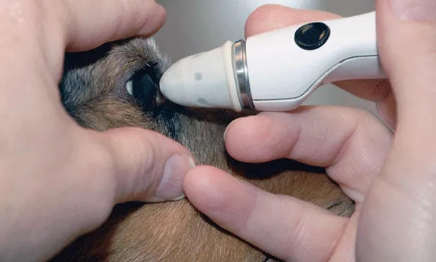
Step-by-Step: Tono-Pen
Step 1
Apply topical anesthetic to the cornea.
Author Insight
IOP determination is best performed in a quiet environment with the animal manually restrained on an examination table.

Step 2
Apply a sterile protective tip cover by gently (2A) placing over the tip and (2B) advancing halfway, (2C) rolling the protective tip down so that the edge rests in the groove at the base of the sensor tip, and (2D) removing the application holder.


Step 3
The cover should (3A) sit smoothly over the applanation tip, but not be so tight as to restrict the movement of the (3B, arrow) sensor tip.
Author Insight
The Tono-Pen sensor must contact the axial cornea directly (ie, not at an angle), thus applanating the cornea at the sensor tip. This is done using a rapid and gentle touch that is repeated.


Step 4
Calibrate according to manufacturer instructions. The indicator window will read Good if the unit calibrates correctly.

Step 5
Avoid excessive globe compression by the hand elevating the superior eyelid and avoid excessive pressure on the neck by the individual restraining the patient; such external pressure may falsely elevate the IOP reading. Obtain readings until the Tono-Pen’s audible tone changes, indicating that 3 readings have been accepted and averaged.

Step-by-Step: TonoVet
Step 1
Insert a new (arrow) rebound tip in the hand piece, and activate the hand piece according to manufacturer instructions.
Author Insight
Because of the limited corneal contact, topical anesthetic is not required for the TonoVet.

Step 2
Hold the rebound probe slightly in front of the cornea and parallel to the floor with thumb on the lower button.
Author Insight
The rebound probe launched from the TonoVet contacts the cornea and rebounds back into the instrument.

Step 3
Press the lower button. Repeat until the TonoVet’s audible tone changes, indicating that 3 readings have been accepted and averaged.
