Dental Diagnoses & Treatment Recommendations
Brook A. Niemiec, DVM, DAVDC, DEVDC, FAVD, Veterinary Dental Specialties & Oral Surgery, San Diego, California
Introduction
Drill down to sharpen your dental skills for common patient problems; carefully studying these images will help you make correct diagnoses and recommend the appropriate treatment option.
Image Gallery

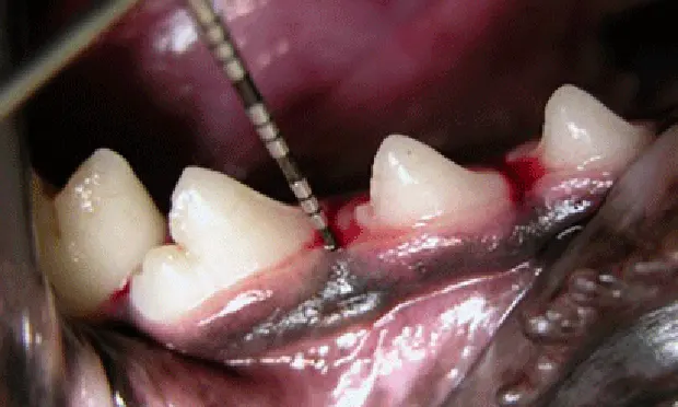
FIGURE 1
Dental radiograph of the left mandibular premolar teeth in a dog showing a classic example of canine tooth resorption.
Diagnosis: When tooth resorption (TR) is seen on routine dental radiographs, the next step is to carefully evaluate the tooth with a dental explorer along the gingival margin and look for clinical evidence of resorption (a sticky or rough area at or just below the gum line).
Treatment: If clinical evidence of resorption is found, the tooth should be extracted. If there is no clinical evidence of a lesion, radiographic monitoring is acceptable, as root resorption is reported to be non-painful in humans.
Extractions of these teeth can be challenging due to the high degree of ankyloses and resorption, and referral to a veterinary dentist is strongly recommended. Crown amputation is not a recognized treatment for these lesions; however, the first premolar tooth (red arrow) may be a candidate, as the end of the root appears isolated from the rest of the tooth and completely surrounded by new bone.