Delivering Supplemental Oxygen
Megan Brashear, CVT, VTS (ECC), VCA Northwest Veterinary Specialists, Clackamas, Oregon
Tissues need oxygen to function.1 Decreased oxygen concentration in circulating arterial blood is known as hypoxemia and may be caused by trauma, disease, and anesthesia.
When hypoxemia is present, respiratory and heart rates increase to compensate for the decreased availability of oxygen. Respiratory effort also increases and provides a subjective measurement of worsening respiration. (See Common Causes of Hypoxia.)
Mucous membrane color provides general information about oxygenation. Pink mucous membranes suggest appropriate oxygenation and perfusion, whereas cyanotic (ie, dusky or blue) mucous membranes indicate decreased blood oxygen concentration. Cyanotic mucous membranes are not always present in patients with hypoxemia, so cyanosis should not be used as an early indicator of decreased oxygenation.2
Arterial oxygen concentration is determined by hemoglobin concentration and the ability of hemoglobin to bind with oxygen molecules. Hemoglobin saturated with oxygen is called oxyhemoglobin. Pulse oximetry (SpO2) evaluates oxygenation using light waves to measure the percentage of oxyhemoglobin in circulating blood. In healthy patients breathing room-temperature air, SpO2 level is 95%.2 Accurate SpO2 results may be difficult to obtain in patients that resent probe placement or those with icteric mucous membranes or poor perfusion. Low ambient light and errors in pulse oximetry usage can alter results.3
Accuracy of readings decreases dramatically when arterial oxyhemoglobin values are below 80%. Supplemental oxygen therapy should be considered in patients with SpO2 below 93% when breathing room air.4
Common Causes of Hypoxia5
Supplemental Oxygen
Supplemental oxygen can be provided using basic equipment or with oxygen cages or chambers, depending on availability and patient need. Use of flow-by oxygen is simple and minimally stressful. Oxygen is set to a flow rate of 3 to 5 L/min and tubing is held 2 to 4 cm from the patient’s face with or without a mask. Some patients may tolerate a mask over their face, which will increase the oxygen concentration, but use caution so the patient is not stressed needlessly. Many patients will only tolerate the oxygen tubing held to their nose and mouth. Delivering oxygen with flow-by methods does not raise the patient’s inspired oxygen concentration markedly, but a patient in respiratory distress can be kept comfortable during physical examination or brief procedures.3
Related ArticlesPlacement of Nasal Oxygen CathetersTop 3 Tips for Intubating Brachycephalic Dog BreedsAssembling a Crash Cart
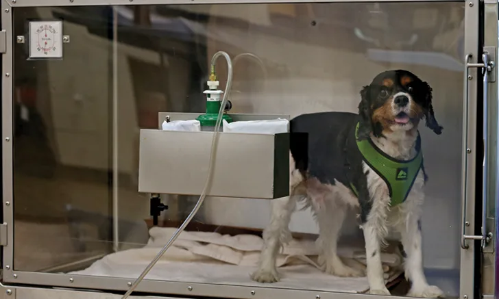
Oxygen Cages or Chambers
Use oxygen cages or chambers to provide oxygen to patients requiring a consistent delivery rate. These special cages can automatically regulate the inspired oxygen percentage and regulate the cage temperature and humidity. They work well for long-term oxygen needs but can be prohibitively expensive for many veterinary practices.

An oxygen hood created using an Elizabethan collar and plastic wrap can supply long-term oxygen support to any patient. Figures courtesy of Megan Brashear, CVT, VTS (ECC)
If an oxygen cage is not available, an oxygen hood can be constructed using plastic wrap, an Elizabethan collar, and oxygen tubing. (See Figure 1.) Place an appropriately sized Elizabethan collar around the patient’s neck and cover two-thirds of the front of the collar with plastic wrap. Make sure to leave a portion of the collar uncovered to allow CO2, heat, and moisture to escape from the improvised hood. Pass the oxygen tubing through the neck of the collar and secure it to the inside using tape. Set the oxygen delivery rate to 1 L/10 kg of body weight to ensure the patient inspires an adequate fraction of inspired oxygen. Monitor patients for overheating and for condensation buildup on the plastic wrap.3 The patient must be under direct observation while the hood is left in place. Patients requiring oxygen support should not be left alone overnight without observation because they may overheat in the hood or dislodge the oxygen source or their condition may worsen. Any animal needing continuous oxygen support should be under direct observation by the veterinary team.
Nasal Oxygen Cannulas
Nasal oxygen cannulas are simple to place and can be used to provide oxygen supplementation for extended periods of time.6 A single cannula can be placed in one naris, or bilateral cannulas can be used. The severity of the patient’s condition and the amount of oxygen he or she needs delivered to improve SpO2 determines whether a single cannula is sufficient.

A bubble humidifier is necessary to humidify oxygen used for long term supplementation.
Oxygen delivered via cannula can dry out nasal tissues and should be humidified via a bubble humidifier connected to the oxygen source and tubing. (See Figure 2.) In cats and small dogs, use 3.5 to 5 Fr red rubber catheters. Large dogs can tolerate 8 to 10 Fr red rubber catheters. (See Figure 3.) A single nasal cannula should deliver 50 to 100 mL/kg/min of humidified oxygen. Bilateral cannulas can deliver 100 to 200 mL/kg/min of humidified oxygen.3

Measuring a nasal oxygen cannula before catheter placement
Patient Monitoring
Patients with respiratory disease requiring supplemental oxygen must be closely monitored for improvement or worsening of their condition. The veterinary nursing team should monitor heart rate, respiratory rate, respiratory effort, and mucous membrane color at least q2h. SpO2 measurements can be used to monitor improvement on oxygen support. The gold standard for monitoring respiratory function is an arterial blood gas, which is not commonly available in most practices, but does not replace careful monitoring by the veterinary nursing team even when available.
Conclusion
Patients with hypoxemia have increased respiratory and heart rates, and mucous membranes may be cyanotic. Hypoxemia is confirmed using pulse oximetry to measure SpO2 and through measurement of arterial blood gas. Most veterinary practices can provide oxygen for short periods of time using simple equipment, and long-term oxygen support can be achieved using an improvised oxygen hood, cannulas, or oxygen cages.