Common Oral Tumors in Dogs & Cats
Tumors can have different appearances and require different treatments. Can you recognize these common oral tumors?
Related Article: Common Masses in Dogs & Cats

Figure 1Figure 1A (above) An oral melanoma in the maxilla of a dog. This dog was treated with maxillectomy and carboplatin chemotherapy.
Figure 1B (left) Appearance of the dog in Figure 1A 2 weeks after maxillectomy. Note the medialization of the lip as a result of using the labial mucosal-submucosal flap for reconstruction of the intraoral defect.
Prognosis: Median survival time is 4–12 months with surgery alone or radiation therapy alone, but may be longer when surgery is combined with either chemotherapy or immunotherapy. The prognosis is highly dependent on clinical stage of disease. This dog survived 7 months.

Figure 2Oral fibrosarcoma in a cat. This cat could no longer eat because of the size of the mass and was euthanized.
Related Article: Surgical Outcomes for Canine Oral Fibrosarcoma
Figure 3Canine oral squamous cell carcinoma (SCC) in the maxilla of a dog. This dog was treated with maxillectomy.
Prognosis: Median survival time is 9–26 months with surgery alone. The addition of radiation therapy or chemotherapy does not seem to improve prognosis. This particular tumor was small and completely excised. This dog lived for 18 months before dying of causes unrelated to the tumor .

Figure 4Oral SCC in a dog treated with segmental mandibulectomy.
Prognosis: Reported median survival, 26–36 months.

Figure 5Oral melanoma treated with mandibulectomy and chemotherapy.
Prognosis: Median survival time is 4–12 months but highly dependent on stage of disease. This patient survived 6 months.
Figure 6Acanthomatous ameloblastoma in the rostral maxilla of a dog treated with a unilateral rostral maxillectomy. These are benign tumors that invade bone, so the prognosis is excellent following complete surgical excision with local recurrence rates <5% and no reports of metastasis.
Prognosis: Reported overall median survival is 64 months. Cure is frequently achieved when clean margins are obtained. This dog did not develop local recurrence.

Figure 7Oral fibrosarcoma in the hard palate and caudal maxilla of a dog treated with combined maxillectomy/orbitectomy.
Prognosis: Median survival times are 7–12 months with surgery alone. Local recurrence is reported in up to 57% of dogs. When treated with a combination of surgery and radiation therapy, the median survival time for dogs with oral fibrosarcoma increases to 18–26 months.

Figure 8Oral SCC in the mandible of a cat treated with mandibulectomy.
Prognosis: Median survival time following mandibulectomy alone is 14 months.

Figure 9Oral Fibrosarcoma on lip; treated with full-thickness lip resection and reconstruction.
Prognosis: Reported median survival, 16–36 months. This patient had incomplete margins and experienced local recurrence in 7 months.
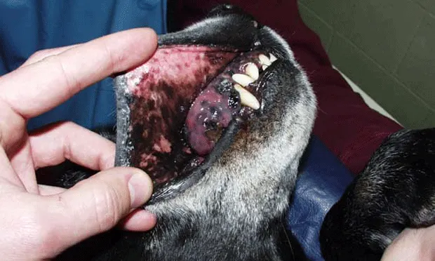
Figure 10Malignant melanoma in the tongue of a dog treated with palliative radiation therapy and gastrostomy tube.
Prognosis: The median survival time for malignant melanoma of the tongue has not been reported. This patient had few treatment options and was euthanized 8 weeks following diagnosis.