Common Ophthalmic Neoplasms in Dogs & Cats
Georgina M. Newbold, DVM, DACVO, The Ohio State University
Diane Van Horn Hendrix, DVM, DACVO, University of Tennessee, Knoxville
Neoplastic lesions can affect every tissue of the eye in both dogs and cats and can occur in the orbit, adnexal tissues (eg, eyelids, conjunctiva), and uveal and retinal tissues inside the globe.1 This article contains a brief overview of commonly encountered ophthalmic neoplasms in dogs and cats.
Orbital Tumors
Orbital masses can cause exophthalmia with decreased ocular retropulsion, strabismus, periocular swelling, and elevation of the third eyelid (Figure 1, below). Although nonneoplastic processes (eg, cellulitis, salivary cysts) may occur, many orbital masses are neoplastic, with a reported frequency of 57.6%.2 Patients with cellulitis, such as that seen with a foreign body or orbital abscess, may present acutely and appear painful, whereas patients with neoplasia may have a more chronic history, typically with little discomfort until later stages of the disease process.

FIGURE 1
Marked exophthalmia with dorsolateral deviation of the globe, periocular swelling, and elevation of the third eyelid in an 8-year-old spayed Vizsla with adenocarcinoma of the right orbit
Common orbital neoplasms include carcinomas (eg, squamous cell carcinoma, adenocarcinoma), sarcomas (eg, osteosarcoma, fibrosarcoma), meningioma, and lymphoma.2 Tumors may extend from local tissues (eg, sinonasal cavities) or originate from the bony or soft tissue structures of the orbit; metastasis from distant sites occurs less often. Orbital mass diagnosis can be confirmed using a combination of imaging (eg, ultrasonography, CT, MRI) and biopsy sampling of associated tissues (Figure 2). Bony lysis is a common finding with many neoplasms.
Adnexal Tumors
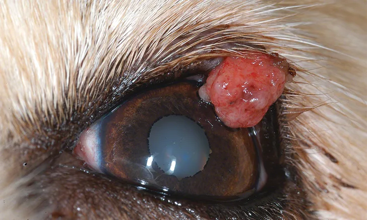
Meibomian gland adenomas (Figure 3) are the most common eyelid tumor in dogs; in cats, squamous cell carcinoma is most prevalent (Figure 4).1,3 Other tumors that affect the eyelid or conjunctival tissues include papilloma (Figure 5), melanoma, hemangioma or hemangiosarcoma (Figure 6), and mast cell tumors (Figure 7). Adnexal tumors in dogs tend to be benign, whereas many eyelid neoplasms in cats have more malignant behavior.1,3

FIGURE 3
Meibomian gland adenoma on the left upper eyelid of a 14-year-old spayed crossbreed dog. Excision was curative.
Tumors of the Globe
Limbal melanocytomas (Figure 8) are benign, heavily pigmented lesions that occur most commonly in German shepherd dogs and Labrador retrievers.4,5 These tumors can expand locally but do not invade the globe or metastasize. They are treated via surgical debulking with diode laser ablation.4
The most common intraocular tumor in both cats and dogs is primary uveal melanoma (Figure 9).6,7 Although these tumors frequently exhibit nonmetastatic but potentially locally invasive behavior in dogs, a greater potential for metastasis is present in cats with iridal melanoma (Figure 10). Surgical removal of the globe is curative if metastasis has not occurred.
Other primary intraocular tumors include iridociliary body adenomas (Figure 11) and medulloepitheliomas. Lymphoma is the most common secondary metastatic neoplasia seen in the eye (Figure 12).1,7 Lymphoma can present with anterior uveitis, chorioretinitis, panuveitis, diffuse iridal thickening, or a discrete uveal mass. Although peripheral lymphadenopathy or other systemic signs are often seen with multicentric lymphoma, ocular signs may be the initial presenting clinical sign.

FIGURE 8
Limbal melanocytoma at the dorsotemporal aspect of the right eye of a 9-year-old spayed crossbreed dog. Because these benign lesions typically progress slowly, the owners elected to monitor rather than treat the lesion.