Colonic Torsion in a Dog
An 8-year-old castrated Siberian husky mixed-breed presented for vomiting and depression in the morning. The dog was 5% dehydrated, tachycardic, and painful on abdominal palpation.
Related Article: Imaging Intestinal Obstruction
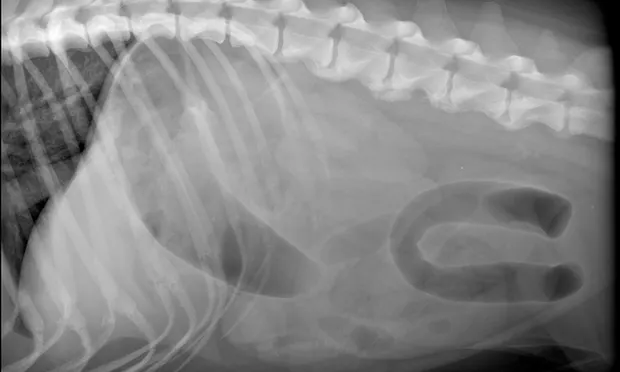
Abdominal radiographs (See Figure 1 above, right lateral; See Figure 2 below, ventrodorsal) showing severe gas distention of the colon (C) with displacement right of midline; there is also fluid and gas distention of the small intestines (SM). When radiographically assessing the intestinal tract, it is important to differentiate colon from small intestine. In this case, colonic torsion was suspected because of the segment of extreme dilation and lack of identification of normal colon.
Results of CBC and serum biochemistry profile were unremarkable. Abdominal radiographs (See Figures 1 and 2) showed a severely gas-distended intestinal segment in the cranial to middle abdomen; other loops of bowel were mildly distended. The colon was not identified in its normal location. Intestinal obstruction from intestinal torsion, possible foreign body, or intussusception was suspected.
Severe fluid- and gas-distended intestines were identified on ultrasound, but a cause of obstruction was not identified. The patient was treated with crystalloid fluid therapy and exploratory laparotomy was performed. The colon, from the level of the ileocolic junction to the pelvic inlet, was severely distended and rotated approximately 90°. Partial torsion of the colon was diagnosed; the cause was undetermined.
The affected bowel remained distended and had diminished tone. Vascular supply to the segment had not been permanently damaged; thus, resection and anastomosis were not pursued.

Colopexy was performed by suturing the unaffected descending colon to the left peritoneal wall using nonabsorbable suture and a muscle-flap technique. Prophylactic gastropexy was also performed using a muscle-flap technique. The abdomen was lavaged with warm saline before closure. The patient had postsurgical diarrhea for 2 days but was clinically normal at suture removal 2 weeks later.
Related Article: Acute Gastric Dilatation-Volvulus in Dogs
A Note on Torsion
Torsion is a twist; volvulus is torsion of an intestine causing obstruction. Torsions of the small intestine, colon, or mesentery are rare but potentially fatal. Presenting signs can include abdominal pain, depression, tenesmus, hematochezia, diarrhea, vomiting, anorexia, and hypovolemic shock. The imaging finding of segmental or diffuse-but-severe intestinal distention, rapid progression of clinical signs, and shock should prompt suspicion of torsion. Early recognition and surgical exploration are important in dogs suspected to have intestinal torsion, as prolonged vascular compromise can result in tissue necrosis, bowel rupture, sepsis, and death. Torsion correction can result in reperfusion injury and shock. Resecting necrotic tissue without attempting to untwist the torsed portion is recommended. Colonic torsions have been reported in collies and German shepherd dogs.1-4
JEAN K. REICHLE, DVM, MS, DACVR, practices at Animal Specialty and Emergency Center in West Los Angeles. Her clinical interests include small mammal elbow dysplasia and imaging. After a rotating internship at VCA West Los Angeles Animal Hospital, Dr. Reichle completed a residency in radiology and earned an MS in radiological health sciences at Colorado State University, where she was assistant professor for 3 years. She earned her DVM from The Ohio State University.
COLONIC TORSION IN A DOG • Jean K. Reichle
References
1. Torsion of the colon in a rough collie. Marks A. Vet Rec 118:400, 1986.2. A case report of torsion of the descending colon in a six-month-old female collie. Endres WA, Remondini DJ, Graver ER. Vet Med Small Anim Clin 63:954-960, 1968.3. Torsion and volvulus of the transverse and descending colon in a German shepherd dog. Halfacree ZJ, Beck AL, Lee KCL, Lipscomb VJ. J Small Anim Pract 47:468-470, 2006.4. Mesenteric torsion in the dog: Two cases. Parker WM, Presnell KR. Can Vet J 13:283-284, 1972.