Colic in the Adult Horse
Colic is the most common reason that horses require emergency veterinarian care. It is known to be the deadliest condition of horses. Colic refers to signs associated with abdominal pain: pawing the ground, flank watching, kicking at the abdomen, stretching, and rolling (Figure 1). GI problems are the most common cause of colic; however, other abdominal organs can be a source of colic (eg, uterine torsion in pregnant mares). Signs of recumbency and inappetence may also be observed, but these are nonspecific to colic.
The first step in assessing the patient is to confirm that the signs are consistent with colic and that there is not another disease process.1 The majority of horses with colic recover with minimal to no treatment. Gas (ie, idiopathic) colic is very common and diagnosed based on a rapid response to analgesia. In one study, 75% of horses with colic recovered with either no treatment or a single dose of an analgesic.2 Fewer than 5% of horses with colic require surgical treatment.

The specific signs associated with colic include pawing at the ground, flank watching, kicking at their abdomen, stretching, and rolling. Photo courtesy of Colorado State University.
Patient Evaluation
Signalment and history
An accurate history is important when evaluating the horses with colic,1 and differential diagnoses will vary based on the age, gender, and breed of the horse. For example, a strangulating pedunculated lipoma should be high on the differential list for older horses, while newborn foals may suffer from meconium impaction.
Because it is common for horses to change owners, determining the duration of ownership is important. Information to acquire from the caregiver includes:
Specific signs shown by the horse
Duration of signs
Previous colic or colic surgery (frequency of or previous colic and surgical history may help identify the cause)
Feeding regimen, as particular feeds (eg, high-grain diets) may be associated with colic
Water source (unheated water sources may freeze in winter, predisposing horses to colic)
Recent change in feeding, stabling, exercise, or travel
Any changes in defecation or urination
Reproductive history should be ascertained for mares
Physical Examination
Owners may misinterpret clinical signs and the clinical condition may change rapidly. Therefore, several observations should be made during the initial approach, allowing the veterinarian time to gain important information regarding the colic diagnosis and severity1:
General attitude (ie, bright, alert, responsive vs dull)
Body condition
Indications of sweating
Abdominal distention
Abrasions to the head, limbs, and tuber coxae which are generally an indication of severe pain
Signs of pain that can be classified as mild, moderate, or severe:
Mild colic signs include intermittent flank staring, kicking at the abdomen, inappetence, lying down, and occasional rolling.
Moderate signs include more persistent rolling with the ability to be distracted; the horse remains standing when walked.
Severe signs include persistent rolling and thrashing with difficulty standing when walking; the horse is generally covered in sweat and often has multiple abrasions to its head, tuber coxae, and limbs.
Cardiovascular status should initially be evaluated and involves examining heart rate (normal,30–44 bpm [adult]), oral mucous membranes (normal, pink and moist with a capillary refill time <2 seconds), extremity temperature, and jugular refill time.
Rectal temperature should be taken (normal, 98.5-101.5°F [adult]); it is unusual for a horse with surgical colic to be febrile. Elevated rectal temperature may be suggestive of colitis, enteritis, or peritonitis, or may indicate another underlying disease process (eg, respiratory tract infection)
The abdomen should be auscultated in all 4 quadrants for borborygmi; absence of borborygmi may be an indication of a surgical lesion.3
Respiratory rate and effort is generally an indication of pain; elevated respiratory rate and significant effort can be observed in horses with severe septic shock.
Abdominal palpation per rectum4 can determine the site of a lesion and help make a definitive diagnosis (use caution to avoid rectal tears and injury).
Safety is imperative; referral is warranted if the veterinary professional does not have appropriate restraint (stocks, experienced handler) or if patient is fractious.
Some basic structures/lesions that can be palpated include:
Dilated small intestine indicative of a small intestinal obstruction
Distended large colon associated with large colon tympany, displacement, or volvulus
Ileal, cecal, pelvic flexure, or small colon impaction
Nephrosplenic entrapment of the large colon
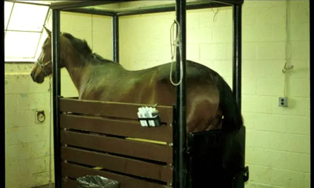
Passage of a nasogastric tube is important, as horses with colic are incapable of vomiting and are at risk for gastric rupture if there is a small intestinal obstruction.
The tube is passed through the nares into the esophagus. A siphon is created using about 1 L of warm tap water. Siphoning is repeated several times to ensure that any gastric accumulation is relieved. The volume of net reflux is calculated; reflux may be suggestive of a small intestinal obstruction.
Diagnosis
Differential diagnosis
The most common cause of colic is undiagnosed or “gas colic.”5 The second most common cause is large colon impaction which most often occurs at the pelvic flexure. Differential diagnoses vary based on geographic region, signalment, and colic severity
Definitive diagnosis
Definitive presurgical diagnosis of colic is not made in most cases. In one study, a definitive diagnosis was not made in 81% of colic episodes.1
Diagnostic tests
Depending on the severity of the colic, laboratory tests may be performed on initial examination. Plasma electrolytes and lactate concentration may be measured with point-of-care tests; PCV and total protein can assess hydration status. Abdominocentesis with peritoneal fluid analysis can be performed. Abdominal radiographs are generally unrewarding in the adult horse except in specific clinical cases; transabdominal ultrasonography can identify dilated loops of small intestine, evaluate peritoneal fluid, and diagnose nephrosplenic entrapment and intussusception.6
Treatment
Medical therapy
Flunixin meglumine (Banamine 1 mg/kg IV [eg, 500 mg or 10 mL for a 500-kg horse]) is an NSAID that is generally the first analgesic administered to the horse with colic. Administration of flunixin meglumine intramuscularly or SC is not recommended, as severe life-threatening clostridial myositis may occur. An injectable form of firocoxib (Equioxx 0.27 mg/kg IV loading dose or 0.09 mg/kg IV maintenance dose [eg, 45 mg or 2 mL for a 500-kg horse]), a COX-1 sparing NSAID, has been released and may be an alternative to flunixin meglumine. Veterinarians in non-US countries typically use hyoscine butylbromide (Buscopan) in the initial treatment of mild colic.
Most horses will respond to single treatment with an NSAID. Occasionally, signs of colic will persist or recur. Xylazine (Rompun 150 mg [1.5 mL] for a 500-kg horse) ± butorphanol (Torbugesic 5–10 mg [0.5 –1 mL] for a 500-kg horse) IV can be administered and should last for 1 hour. Horses that are violently painful should be sedated with detomidine (Dormosedan 5-10 mg [0.5-1 mL] IV for a 500-kg horse).
Horses with colic may also benefit from medication administration through a nasogastric tube with water/electrolytes/mineral oil (typical volumes of 4-6 L). The tube should be in the stomach prior to administration of mineral oil or water/mineral oil. A funnel is generally used to slowly administer the water or mineral oil. It is important to avoid getting any mineral oil in the nasopharynx/larynx or trachea. Aspiration of mineral oil is associated with almost 100% fatality.
The horse should always be checked for reflux prior to nasogastric tube fluid administration to prevent gastric rupture. Horses that have nasogastric reflux or small intestinal distention on rectal palpation should not be treated with fluids via the nasogastric tube.
IV fluids may also be given; however, horses are generally referred to a hospital if IV fluids are necessary. Adult horses are often given a 10-20 L IV bolus of polyionic isotonic crystalloid fluids. Maintenance fluid rates are 2 mL/kg/hour, about 1 L/hour for a 500 kg horse.
It is recommended to withhold feed and water while the horse shows signs of colic. When the colic resolves, the horse is started back on a reduced amount of feed and water. If tolerated, the amount is increased over the following days until normalized.
Why & When to Refer
The main indication for referral is severe or persistent abdominal pain. As a guideline, if the horse has not responded to a dose of flunixin meglumine followed by xylazine (± butorphanol) then referral to a surgical facility, as well as estimated costs, should be discussed with the owner.
Other indications for referral include nasogastric reflux, shock (tachycardia, toxic oral mucous membranes, cool extremities, poor jugular refill), cecal or small colon impaction on palpation per rectum, moderate to severe abdominal distention, lack of fecal production, and persistent absence of intestinal borborygmi.
Occasionally owners are unwilling to pursue further diagnostic tests and treatment at a referral hospital for financial reasons. At-home treatment includes analgesia and supportive IV fluid therapy.
Indications for euthanasia are abdominal pain that is unresponsive to analgesia, abdominal pain that persists and recurs following repeated doses of analgesia (eg, beyond 24–36 hours), cardiovascular deterioration (ie, increasing heart rate, development of injected or toxic mucous membranes, coolness of extremities), and severe or increasing abdominal distention. Abdominal ultrasonographic examination6 can be used to identify an increase in peritoneal fluid volume or distended loops of small intestine. Abdominocentesis can also be performed; serosanguineous fluids may indicate intestinal ischemia and ingesta may indicate intestinal rupture/enterocentsis.
Surgery
Abdominal exploratory surgery is a diagnostic as well as a therapeutic procedure for colic and is performed at a surgical referral hospital by a board-certified veterinary surgeon (DACVS), and is indicated on an emergency basis in persistently or severely painful horses. Often a definitive diagnosis is not made before surgery. Early referral and surgical treatment are major contributors to improved survival. The prognosis for survival following colic surgery is good to excellent for most horses.
Follow-Up
Most horses with colic recover with minimal to no treatment. It is important to follow up with caregivers following initial treatment to ensure that the signs are not persisting and that the horse has returned to normal demeanor, activity, appetite, and fecal production.
Species at a Glance
Colic is the most common reason that horses require emergency veterinary care.
Most horses with colic recover with minimal or no treatment.
Horses showing signs of colic with a high heart rate (>60 beats/minute) should have a nasogastric tube passed immediately because it can be an indication of impending gastric rupture.
Persistent or severe pain is the most important indication for referral.