CO2 Lasers & Radiosurgery: What’s the Difference?
Over the past 10 years, there has been a dramatic increase in advanced technologies and their use in veterinary surgery.
New surgical devices such as CO2 lasers and radiosurgery units have been readily accepted by practitioners.1-5 These devices are no longer restricted to the referral specialist and many private practices incorporate use of CO2 lasers or radiosurgery units. Unfortunately, much of the marketing, lay publications, and conference presentations surrounding these instruments have been based on anecdote and opinion rather than science.
How They Work
All CO2 lasers and radiosurgery units cause cellular destruction in the same way-by vaporizing intracellular water. Although they deliver energy to tissue in different ways, the histologic characteristics of the incisions are essentially the same-an immediate area of vaporization, surrounded by a zone of irreversible thermal or coagulative necrosis, and a further zone of reversible edema and inflammation.
Radiosurgery
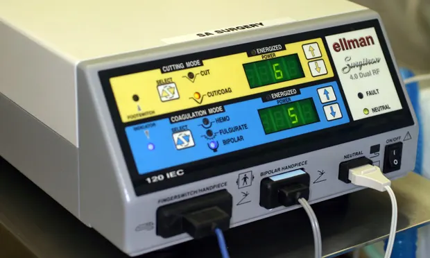
Radiosurgery uses electricity to produce ultrahigh radiofrequencies (≥ 3.8 MHz) that agitate water molecules to the point of vaporization. Radiosurgery units offer monopolar and bipolar applications (controlled by foot pedal and finger switch), which allow use of several different waveforms:
Pure filtered waveform for cutting
Fully rectified waveform for blended cutting with coagulation
Partially rectified waveform for coagulation
Fulguration for ablation
Pinpoint bipolar coagulation.
There is a wide variety of soft tissue, dental, microsurgical and endoscopic instruments (wands/tips) available for use with radiosurgery, which makes it versatile for a wide range of veterinary applications (Figure 1). Radiosurgery should not be confused with electrosurgery, which operates at much lower frequencies and causes considerably greater collateral damage.
Laser Surgery
LASER (Light Amplification by the Stimulated Emission of Radiation) surgery relies upon production of electromagnetic radiation in response to photon emission by the lasing medium.6 The lasing medium most commonly exploited in veterinary medicine is CO2 (10,600 nm).
The laser beam is activated by foot pedal and transferred from the base unit to the ceramic handpiece by either an articulated arm or a flexible waveguide (Figure 2), with the surgeon able to control power and waveform, which consist of:
Continuous laser
Pulse with laser energy supplied as high-amplitude, short duration pulses, followed by short pauses during which tissue is allowed to cool thereby reducing damage
Superpulse with frequencies around 200 per second to permit primary incisions while maintaining lower tissue temperatures and less damage.
The characteristics of the laser beam as it emerges from the ceramic handpiece can be controlled in 2 ways-size of the ceramic tip (0.25 to 1.0 mm) and whether the beam is focused (cutting) or defocused (ablation). The size of the ceramic tip determines the diameter of the laser beam and, therefore, the amount of tissue destroyed. Finer tips are used for incising, while larger tips are preferred for vascular tissue and ablation.
Advantages & Disadvantages
Technique & Collateral Damage
With proper technique (See Suggested Reading in Aids & Resources), collateral damage and associated inflammation is lower with both small-spot focused CO2 lasers and microfiber radiosurgery set to filter-cut mode than with cryosurgery, electrosurgery, or diode laser. Ultrafine incisions also permit delicate and precise dissection with less postoperative pain and faster healing with less scarring (Figures 3 and 4). However, the greater collateral damage created by diode lasers does enable them to seal larger vessels (up to 2 mm in diameter).
CO2 laser handpieces and ceramic probes do not contact the tissue and, therefore, there is no tissue drag, but a lack of tactile feedback; a learning curve is needed to master the technique. In addition, since there is no contact, no charred tissue collects on the laser tip. Radiosurgery requires tissue contact and provides tactile feedback similar to scalpel blade incisions, but tissue drag may become a problem unless the electrode is kept clean.
Scientific Studies
Studies objectively assessing different surgical cutting modalities have generally done so by comparison to a gold standard that causes little to no collateral damage (ie, scalpel blade). Unlike electrosurgery, radiosurgery and CO2 lasers offer superior accuracy and reduced collateral damage.
Histological studies involving human skin peel grafts concluded that radiosurgery produces less epidermal loss, reduced zone of thermal damage, and reduced dermal fibrosis than CO2 laser.7 In other comparative studies including turbinate ablation and resection, biopsy collection, and effects on oviductal tissue, radiosurgery was comparable or superior to CO2 laser.8-10
In veterinary medicine few comparative studies have been published; however, results in greyhounds, pigeons, and green iguanas, suggest that radiosurgery is superior to CO2 laser.11-13 To date, there have been no veterinary studies to assess the effects of laser or radiosurgery on healing. However, a human study compared radiosurgical and CO2 laser resection of nasal turbinates and concluded that although both methodologies were equally effective at accomplishing the surgical goals and resolving nasal obstruction, CO2 laser resulted in significantly greater disturbance of mucociliary function.14
Postoperative Pain
There have been few objective comparisons between devices in regard to postoperative pain. Nevertheless, two independent studies involving soft palate resection in humans provide convincing evidence that radiosurgery produces less postoperative pain than CO2 laser surgery.10,15 Furthermore, postoperative side effects (trouble with smell and taste, pharyngeal dryness, globus sensation, voice change, and pharyngonasal reflux) and complications (wound infection, dehiscence, and posterior pillar narrowing) were more common following CO2 laser surgery.10
Health & Safety Concerns
There are several significant health and safety issues that must be appreciated and addressed when using CO2 lasers. To prevent retinal damage to personnel in the operating room, protective glasses must be worn at all times lasers are in use.16 Likewise, the patient's eyes should also be protected by gauze or drapes, unless specifically operating on ocular tissues. Windows in the operating room should be covered to prevent potential damage to anyone passing by.
All medical lasers come with emergency cut-off switches that can be operated by those in the room or when the operating room door is opened.16 Both radiosurgical and CO2 laser devices produce a smoke plume that may contain pyrolysis products known to be cytotoxic.17,18 This smoke, which can contain viable bacterial, viral, or patient DNA, should be evacuated from the surgical site by filtered vacuum.
Limitations
Even when using ultrafine cutting techniques, both devices cause similar coagulative damage along the incision. Such artifacts can adversely affect histological interpretations, which is an important consideration if margin evaluation is important. Unlike radiosurgical units, CO2 units cannot be used in a liquid medium and there are few terminal attachments. The Table summarizes comparisons between radiosurgery and CO2 laser units.
Economic Impact
Radiosurgery units ($4300-$14,000) are 3 to 10 times less expensive to purchase than CO2 lasers ($21,000-$33,500). Given their comparable effects, practitioners would do well to try both modalities and determine applications for either before embarking on a major capital purchase. Personal preference, not effectiveness, appears to be the factor in determining which unit to choose.
Dr. Divers discloses that he has participated in research studies funded by Ellman International and Universal Lasers. He does not endorse any particular product or brand.