Clinical Suite: Lumps & Bumps
Barbara E. Kitchell, DVM, PhD, DACVIM (Internal Medicine & Oncology)
Jessica Goodman Lee, CVPM, Veterinary Credit Plans, Irvine, California

Overview
Barbara E. Kitchell, DVM, PhD, DACVIM (Internal Medicine & Oncology)
AN EARLY START
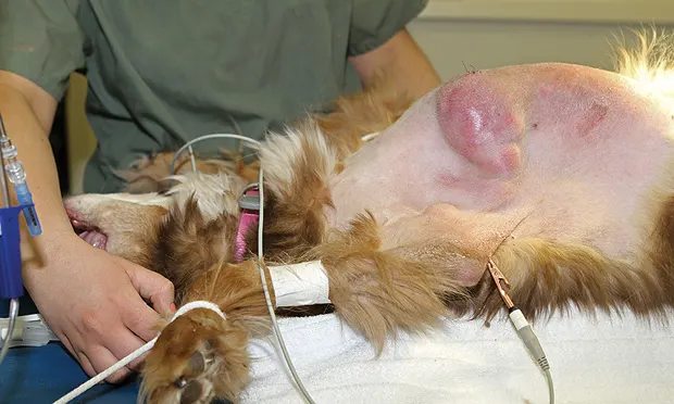
Skin and subcutaneous (SC) tumors are frequently discovered in routine examinations. Dermal and SC tumors are often benign, but there is still potential for malignancy, warranting additional care. Although some tumor types have metastatic potential from their inception, in many cases the potential for a malignant tumor to become invasive and metastatic is linked to its size. For the majority of skin and SC tumors, surgery is the treatment of choice. Early intervention offers the best chance for complete surgical excision and long-term local control. For some tumors, metastasis occurs relatively late in the course of disease, and early local control of the primary tumor can reduce the risk for metastatic disease.
KEY QUESTIONS
The team should acquire as much history as possible from the client:
How long has the mass been present?
Has the mass increased in size? How quickly?
Does the mass bother the patient?
Are there signs of illness?
Are there any changes in the patient’s behavior?
Did the patient suffer any trauma in the mass area?
Has the patient visited an area with known fungal infection risk?
Is the patient up-to-date on vaccinations? Where on the body were the vaccines administered?
The size, shape, and mobility of the mass are of particular interest:
Is the mass symmetrical or irregular?
Is the mass fixed or mobile?
If the mass moves, does it move with or under the skin?
Is the overlying skin intact or ulcerated?
Is there any apparent pain or discomfort?
A complete examination should include lymph node evaluation and a thorough check for other lesions. Measurements of the mass should be taken in 2 dimensions (ie, length and width), with depth as an optional third. Masses should be aspirated and evaluated by cytology; if the result is ambiguous or does not agree with the clinical impression, histopathology is recommended. A common misconception is that biopsy or needle aspiration can result in tumor spread. Although needle-tracking into surrounding tissues is possible, metastasis is not a significant risk from needle aspiration or appropriate biopsy procedures.
Primer
Barbara E. Kitchell, DVM, PhD, DACVIM (Internal Medicine & Oncology)
Tumors originating from dermal and SC tissue can be epithelial, mesenchymal, or round-cell. Benign lesions, common in older dogs, can include lipomas, sebaceous adenomas, epidermal inclusion cysts, papillomas, histiocytomas (especially dogs <3 years of age), and granulomas. In cats, the most common benign skin tumors are basal cell tumors, often localized to the head and neck. Both dogs and cats may have multiple benign masses, so it is important to measure and map each mass for future reference. This makes identifying new lesions or changes in existing ones much easier and can indicate the need for additional diagnostics.
MAST CELL TUMORS
Mast cell tumors (MCTs) are common in dogs and cats.
In cats, cutaneous MCTs are usually benign; in dogs, the biological behavior is more variable.
MCTs can occur in any dog breed but are common in boxers, Boston terriers, pugs, and Labrador retrievers.
MCT size can fluctuate because of degranulation and secondary inflammation (Darier’s sign).
SQUAMOUS CELL CARCINOMA
Squamous cell carcinoma (SCC) is fairly common in dogs and cats.
SCC is usually found in areas lacking pigment or fur.
In cats, these sun-induced tumors can be described as “fight wounds that don’t heal.”
MESENCHYMAL TUMORS
Mesenchymal tumors include a variety of soft tissue sarcomas: fibrosarcomas, hemangiopericytomas, and peripheral nerve sheath tumors.
In cats, soft tissue sarcomas can be associated with SC injections (eg, adjuvanted rabies, feline leukemia vaccines).
The skin lesions of dogs with cutaneous lymphomas can closely mimic inflammatory dermatopathies; biopsy is essential for confirmation.
Sarcomas may require an incisional biopsy for adequate diagnosis.
Biological behavior of sarcomas is best predicted by histologic grade.
NEXT STEPS
After establishing the diagnosis of cutaneous or SC masses, the next step is diagnostic staging.
Recommended staging tests depend on the mass’s biological behavior and the metastatic pattern of the specific tumor type:
This can include thoracic radiography, abdominal ultrasonography, and lymph node cytology or histopathology.
Diagnostic results can help determine the appropriate course of treatment.
Surgery is the most curative modality for cancer therapy.
Radiation therapy can be excellent for local control of skin tumors that cannot be completely surgically excised.
Chemotherapy is used most commonly for tumors that have metastasized or are at high risk for metastasis.
Communication
Jessica Goodman Lee, CVPM Brakke Consulting Dallas, Texas
Finding a lump or bump on our pets or ourselves can be scary and should always be taken seriously. As a team member, regardless of position, it is your responsibility to let clients know the importance of having all lumps and bumps examined by a veterinarian. The key is not to frighten clients, while at the same time being very careful not to minimize the need to have the masses checked. This is also a wonderful opportunity to show how much everyone cares about the patient.
What to say if a mass or growth is found:
It’s important to always have a veterinarian take a look at these. I will make sure to let her know about it.
We always want to make sure the veterinarian knows about any changes since the last time we saw Missy, no matter how small they may seem.
What not to say:
I’m sure it’s nothing.
I wouldn’t worry about it.
Besides veterinary technicians and veterinarians, whose responsibility it is to thoroughly examine the patient and discuss client concerns, the important role that receptionists and grooming teams can play in this process should not be overlooked.

Receptionists
Scenario 1: When a client calls for an appointment
Receptionist:Okay, I have Missy scheduled for an appointment with Dr. Jones on Thursday. One of the things I’d like you to do before then is take a few minutes and give her a good all-over massage. The purpose of this is to see if you feel anything different—such as a growth or lump of any kind.
Owner:No one has ever asked me to do this before. Is there something I should be concerned about?
Receptionist:Not at all. We are recommending all of our clients do this to ensure that nothing is missed. Just as with humans, these things always need to be checked out, and we are working hard to make sure that any changes are detected early. Dr. Jones will do a thorough examination, but often you as the owner are the best source of information!
Owner:That makes sense. What should I do if I find something that I think she should look at?
Receptionist:Before bringing Missy to the veterinarian, jot down on a piece of paper where it is and what it feels like. You can even take a marker and put a little “x” over it to make it easier to find [depending on the pet’s coat].
Scenario 2: When the receptionist can come out from behind the desk to bond with the client and patient in the lobby without neglecting the phone or other clients
Receptionist:Bubba, I forgot how handsome you are…come over here and let me pet you! [Gives the patient attention while at the same time providing a casual “once over” with hands]
Hmmm…what’s this I feel? [Finds a small lump on his back and parts the fur]
Is this something new, or has it been there for a while?
Owner:I noticed that a couple of months ago, but I figured it was nothing—just part of getting older. He’s going to be 12 this year!
Receptionist:And you still take great care of him! I’m just going to make sure Dr. Jones takes a look at this. Are there any other changes that you’ve noticed?
Owner:Actually, I felt another lump just the other day.
Receptionist:If you like, we can grab a marker and put a little “x” on these spots. I’m going to let Dr. Jones know about these, but this way she can find them easily. Would that be okay with you?
Kennel & Grooming Teams
These teams often have the most hands-on interaction with many patients, sometimes on a regular basis. These individuals should receive training on how to perform a head-to-toe body “feel,” create a protocol to document areas of concern, and inform a veterinarian of the findings. This provides a prime opportunity for early detection and patient advocacy. If a patient is boarded or groomed regularly, kennel teams should also take the time to see whether previously found areas of concern are changing (eg, size, texture) and inform the client.
Kennel Attendant:Missy had a lot of fun this time and she ate all of her food!
Owner:I am so glad to hear that! You know how much I hate leaving her, but my travel schedule has just gotten so crazy.
Kennel Attendant:We love having her here—she’s one of my favorites! I spend a lot of time with her, but this time when I was rubbing her belly I noticed a small lump on the left side of her chest. Here, let me show you [can show client in the lobby or examination room]. Have you noticed that before?
Owner:A lump? I’ve never felt anything…oh no, do you think it is serious?
Kennel Attendant:Let’s have Dr. Jones take a look and see what she thinks.
Depending on hospital policy, at this point the kennel attendant should provide further information and follow-up recommendations.
From then on, every time Missy boards, the team should make sure not only to check her for new lumps and bumps but also to keep an eye on any changes to the ones already identified.
Workflow
Jessica Goodman Lee, CVPM Brakke Consulting Dallas, Texas
Technician/Assistant
Escort client & patient to examination room
Obtain medical history
Ask client about any skin abnormalities
Briefly look at any areas noted by client or receptionist
If time allows, take a few minutes to complete quick examination
Document all observations and conversations in medical record
Technician or Receptionist
Schedule upcoming tests, appointments, and rechecks as instructed by veterinarian
Ensure client is comfortable with treatment plan and has received all handouts as recommended by veterinarian
Roles
Jessica Goodman Lee, CVPM Brakke Consulting Dallas, Texas
Receptionist
Role: Initial client point of contact & patient/client bonding expert
Responsibilities:
Whenever possible, take time to interact with patient and feel for any abnormalities on skin
Communicate any abnormalities verbally and in writing to technician/veterinarian
Before appointment, recommend that clients feel pets from head to toe and note any lumps, bumps, or concerns; recommend that client feel the lumps and mark them with a marker to make them easy to locate during examination
Technician
Role: Client educator & pet advocate
Responsibilities:
Take history of masses
Record clinical signs of illness
Point out any new masses to client and veterinarian
Discuss clipping lesion area with client and obtain consent
Clip and clean open or draining lesions as directed by veterinarian
Complete tumor map under clinician direction, including 2- to 3-dimensional caliper measurements of all lesions, for medical record and copied for client
Assist in performing fine-needle aspiration of lesions and prepare cytology slide, or obtain aspiration materials under veterinarian direction (if appropriate)
Be familiar with in-house staining procedures to facilitate diagnosis of benign lesions
Practice Manager
Role: Supervisor of team/client education & practice protocols
Responsibilities:
Market importance of early diagnosis for optimal patient healthcare
Develop brochure about common skin and SC lesions in dogs and cats
Organize individual client handouts on commonly diagnosed masses or growths
Ensure microscope is in good working order for in-house diagnostics
Organize team education seminars and assign veterinarian to create presentation
Training
Jessica Goodman Lee, CVPM Brakke Consulting Dallas, Texas
Creating a practice that emphasizes and encourages learning and participation for everyone on the team is one of the most powerful ways to enhance teamwork, client service, and patient care. It must always be crystal clear that only a veterinarian can make diagnoses, but educating the entire team about veterinary medicine enables everyone to have intelligent and meaningful conversations with clients about their pet’s healthcare. Benefits include increased client bonding opportunities, client education, and compliance.
Tips
Improvements in therapeutic options and protocols warrant semiregular team training sessions.
Remind the team that only the veterinarian can make diagnoses.
BACK TO BASICS
When it comes to lumps and bumps, one of the best ways to educate the practice team is to have a veterinarian create a presentation that covers the basics:Most common types of masses and growths
What they look like
What they feel like
Treatment and surgical options
Signs to look for based on patient age and breed
ROLE PLAY!
Develop scenarios based on the practice’s specific needs and clientele
Focus on the ability to communicate concerns to and elicit important information from clients without frightening them
Educate team members to always put themselves in the client’s shoes
Remember, just the words growth and mass can be really scary for anyone!
Know the sources
Many sources are available to help educate the veterinary team about lumps and bumps, including:
Brief Connection
The following can help veterinarians provide quick answers for the team education seminar:
Handout
Barbara E. Kitchell, DVM, PhD, DACVIM (Internal Medicine & Oncology)
You asked… the expert answers!
Q:
“What is this mass on my pet?”“Could it be cancer?”“How will the veterinarian diagnose the lump?”“What are the options for treatment?”
A:
Many owners who find lumps and bumps may have the same general questions, and it is essential to let your veterinarian know about your discovery as soon as possible. Dogs and cats may have many benign skin tumors, but confirming the diagnosis with laboratory work (eg, cytology or biopsy) is very important. The results of these tests can help your veterinarian detect malignant lesions before they spread.
Even some benign tumors warrant further workups. Benign tumors should be monitored for signs of change, because it is possible for a benign tumor to transform into a malignant tumor.
Malignant tumors can be similar in appearance to benign tumors, so being proactive with detection and treatment (not “waiting and watching”) can help your pet’s health and prognosis.
Download the "You Asked...The Expert Answers" handout
Lumps & Bumps: Steps After Diagnosis

Once the diagnosis of your pet’s lump or bump has been confirmed, the next step is to discuss all treatment options with your veterinarian.
If cancer is detected…
Surgery is the most common (and most effective) curative method for treatment. Talk to your veterinarian about presurgical steps you can take to prepare your pet.
If surgery isn’t an option…
Radiation therapy can be an effective alternative for tumor therapy. Some tumor types require chemotherapy for control.
Remember to keep an eye on things…
Your veterinarian can provide you with a tumor map of the size and location of your pet’s lump(s) or bump(s). This can help you note any changes in the bump’s size and identify any new lesions.
Careful monitoring should help you detect changes early on so you can optimize your companion’s chances at having a long and happy life.