Clinical Challenge: How Artificial Intelligence Can Improve a Lifetime of Care
Sponsored by Zoetis
In-clinic technology, including artificial intelligence (AI) and digital cytology, is advancing quickly in veterinary medicine. AI diagnostic technology can be used for rapid evaluation of fecal samples, blood smears, and dermatologic cytology. Other advances, like digital cytology with remote pathologist interpretation, also allow for real-time test results. Together, these tools can increase the efficiency of the veterinary team, improve patient care, and improve workflows in the clinic.
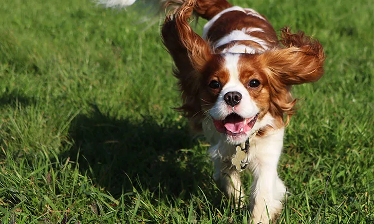
Follow along as Blaze, the Cavalier King Charles Spaniel, benefits from these new diagnostic capabilities, and see how his care team puts them to use in daily practice.
The First Puppy Visit
Blaze, a 10-week-old, male Cavalier King Charles Spaniel, is presented for his initial puppy visit. Along with other recommended care, a fecal sample for parasite testing is approved by his owner. This sample will be processed by the clinic’s new diagnostic tool, the Vetscan Imagyst®.
With the Vetscan Imagyst, veterinary team members are no longer tasked with microscopic evaluation of fecal samples, freeing up time for the support staff to do other tasks and eliminating the risk for human error during sample evaluation.
When using the Vetscan Imagyst AI Fecal application, a fecal sample is processed using the Vetscan Imagyst sample preparation kit. After preparation and centrifugation, a transfer loop is used to transfer a portion of the sample to a slide, and a specialized coverslip is applied.2 The slide is then digitally scanned and uploaded for evaluation by a deep learning algorithm trained by diagnostic parasitology experts.2 The Vetscan Imagyst AI Fecal algorithm provides accurate detection of many GI parasites of dogs and cats, including common species of hookworms, roundworms, whipworms, tapeworms, coccidia, and Giardia (Table).1,2
Due to variability in methods used for fecal flotation and the skill level of the team members reading the sample, it is estimated that parasites are missed in up to 50% of dogs infected with GI parasites when passive flotation is used in private practice.3,4 In addition, some parasites like Giardia shed intermittently or in small numbers, making their detection difficult by traditional flotation methods.5 Based on studies,1,6 the Vetscan Imagyst AI Fecal algorithm is able to correctly identify common GI parasite eggs, cysts, and oocysts in canine and feline fecal samples comparable to manual evaluation by diagnostic parasitology experts (Table).
TABLE: Algorithm performance as compared with manual identification by a diagnostic parasitology expert (at 100%)1,6
*Canine and feline samples combined; **Canine samples only. Trichuris spp ova not seen in feline samples6
Results
In Blaze’s fecal sample, 5 roundworm eggs are detected. An appropriate dewormer is prescribed, and a fecal recheck 7 to 10 days after treatment is recommended to ensure clearance of infection. The veterinary technician provides the owners with a printout that includes images of the roundworm eggs detected as well as general information about roundworms, including their zoonotic potential to humans.7
Time for Neuter
A few months later, Blaze is presented to be neutered. Prior to the procedure, a routine CBC and serum chemistry profile are performed. Serum chemistry is within normal limits, but the automated CBC reveals a decreased platelet count of 70,000/µL. CBC results are verified with a blood smear that is analyzed by the Vetscan Imagyst AI Blood Smear application.
Although review of a blood smear aids interpretation of CBC results, they are not always performed in daily practice due to time and lack of training; the Vetscan Imagyst offers a solution to these hurdles.8 Vetscan Imagyst Al blood smear technology provides a WBC differential, detects platelet clumps, provides estimated platelet counts, and identifies nucleated RBCs and polychromatophils (immature RBCs), which are indicators of regeneration.8 The results are highly accurate and comparable to those of board-certified clinical pathologists.8 In cases in which results are concerning or additional information is needed (ie, identification of blood parasites), review by a clinical pathologist can be added (additional fees may apply).8
Results
Results confirm thrombocytopenia, and very few platelet clumps are detected. Considering the patient’s breed, macrothombocytopenia, an inherited condition present in ≈30% to 50% of Cavalier King Charles Spaniels, is suspected.9 It typically presents with thrombocytopenia and macrothrombocytes but normal coagulation.9 To confirm this suspicion, an add-on expert review by a board-certified clinical pathologist is ordered.
Pathologist review confirms the presence of numerous macrothrombocytes. Surgery is elected, given the high prevalence of inherited macrothrombocytopenia in Cavalier King Charles Spaniels.
Use of the Vetscan Imagyst to review the blood smear allowed verification of CBC results and rapid review by a clinical pathologist. This provided quick peace of mind for the clients and allowed surgery to occur the same day rather than having to postpone and reschedule while waiting for results.
Adult Sick Pet Examination
At 3 years of age, Blaze is presented for headshaking and scratching at his ears and ventral abdomen. On examination, thick, dark brown, waxy exudate with erythema is identified in both ear canals. The tympanum cannot be viewed. The skin in the inguinal region is diffusely erythematous with occasional crusts.
The veterinarian recommends ear and skin cytology to guide treatment. The veterinary team collects and prepares Blaze’s ear swabs and a skin impression smear and scans them into the Vetscan Imagyst for analysis. While the sample is being analyzed, the veterinary team members proceed with client communication.
The Vetscan Imagyst offers AI dermatology cytologic review of ear smears, skin impression smears, and skin swabs.10 Yeast, cocci and rod bacteria, and inflammatory cells can all be detected with accuracy comparable to board-certified clinical pathologists.10 Cytology is a powerful tool in dermatology that can help guide treatment plans and support antimicrobial stewardship by confirming the presence of bacteria and/or yeast prior to the institution of treatment.
Results
Large amounts of yeast, cocci, and inflammatory cells are detected bilaterally with ear cytology. Skin cytology reveals cocci and numerous neutrophils. Otitis externa and pyoderma are diagnosed, and a treatment plan that addresses both pruritus and infection is developed. The client is given a printout containing images of the microorganisms found with cytology, providing a visual representation of the degree of infection and improving the perceived value of the diagnostic testing.
Adult Wellness Examination
At 5 years of age, Blaze comes in for his annual physical examination. During the physical examination, a small (5-mm diameter) dermal nodule near the right hip is identified. Aspiration and cytology of the lump are recommended, and the owner agrees. Aspirates are obtained and slides are prepared, stained, and scanned using the Vetscan Imagyst.
With the Vetscan Imagyst, sample submission can include up to 4 slides for a set cost. Samples from up to 2 sites can be included for the same base cost, as long as the maximum number of slides does not exceed 4.12 If a sample is nondiagnostic, a resubmission is allowed for no additional fee. Consultation with a board-certified clinical pathologist is available as needed.
Results
Blaze’s cytology results are reported in 32 minutes and reveal a diagnosis of a mast cell tumor. A treatment plan is discussed during this visit, and the owner schedules Blaze for surgical excision of the mass. As compared with traditional turnaround times, Blaze’s owner was able to get results during the initial appointment and discuss next steps with his care team without delay.
Conclusion
The Zoetis Vetscan Imagyst provides rapid and accurate fecal testing, blood smear evaluation, dermatologic cytology, and digital cytology through a combination of artificial intelligence and remote evaluation and support by board-certified clinical pathologists.
Over the course of Blaze’s life, he was able to benefit from rapid and accurate diagnostic results, which also allowed his care team to efficiently address his medical needs and provide personalized care to both him and his owner. The Vetscan Imagyst improved efficiency and workflow for his care team, allowing them to spend time where it counts most: with Blaze and his owner.
VTS-01138