Chronic Vomiting in a Dog
Michael E. Matz, DVM, DACVIM, Veterinary Specialty Center of Tucson, Arizona
A 17-month-old, spayed female bullmastiff was evaluated for vomiting of 5 weeks duration.
History. Vomiting initially occurred once per week but increased to twice per day during the 2 weeks before presentation. The vomitus consisted of food, fluid, and bile; vomiting was not consistently associated with eating. Appetite and activity level remained normal, and diarrhea did not occur.
The dog had an unremarkable medical history. Four months previously, she had been presented for a shoulder lameness that rapidly resolved after treatment with carprofen for 2 weeks. The dog had not received any medications recently except monthly milbemycin.
Physical Examination. Physical examination was normal. The referring veterinarian did not identify any abnormalities on CBC, biochemical profile, or fecal examination.
Diagnostic Testing. Abdominal ultrasonography showed nothing abnormal. Upper gastrointestinal endoscopy was done with the dog under general anesthesia. There were blades of fresh grass in the stomach; several mucosal ulcers (3 to 5 mm in diameter) in the gastric body; and many 1- to 2-mm, smooth, reddish lymphoid follicles in the gastric cardia. The duodenal mucosa was slightly granular. Several duodenal and gastric biopsy samples were collected and a duodenal aspirate was negative for Giardia. Figure 4 (below) is a photomicrograph of a gastric cytology sample viewed with oil immersion.
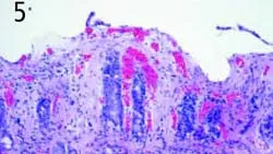
Diagnosis: Moderate lymphoplasma-cellular gastritis with erosion and suppuration
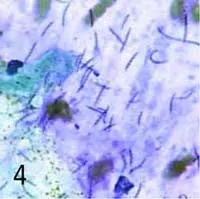
Ask Yourself ...• What technique is used to obtain mucosal brush cytology specimens during endoscopic examination of the stomach?• What are the potential diagnostic benefits of obtaining and evaluating gastric cytology specimens?• What is the significance of the cytology findings in Figure 4? (right)
The histopathologic diagnosis was moderate lymphoplasmacellular gastritis with erosion and suppuration (Figure 5, below). Spiral bacteria were visible with routine stains. The duodenum was normal. The dog was entered into a study assessing two treatments for Helicobacter species and was randomly assigned to receive 15 mg/kg amoxicillin Q 12 H, 10 mg/kg metronidazole Q 12 H, 524 mg Pepto-Bismol (Procter & Gamble) Q 12 H, and 0.5 mg/kg famotidine Q 12 H for 2 weeks.
Four weeks after completion of therapy, gastroscopy was performed. Vomiting had not occurred during the preceding 4 weeks. The gastric ulcers had healed and were not visible, and the lymphoid follicles appeared indistinct. Histopathologic evaluation revealed minimal gastritis, and spiral bacteria were not visible with routine or special stains or detected in gastric cytology specimens. Vomiting occurred only 3 times in the next 5 months.
Although a cause-and-effect relationship between Helicobacter species and chronic gastritis and vomiting has not been proven in dogs and cats, the clinical response of this dog to treatment suggests that Helicobacter species may play a role in some cases. Further investigation is necessary to define the potential role of Helicobacter species in dogs and cats with chronic vomiting. However, presently it seems prudent to use gastroscopy to determine whether gastric spiral bacteria are present in vomiting dogs and cats. Collection and assessment of a gastric cytology specimen is an inexpensive, rapid, and accurate method to identify the bacteria.
Did You Answer...• A guarded cytology brush is passed through the endoscope's biopsy channel while the endoscope is positioned within the gastric body (or adjacent to the area to be sampled). The brush is extended from the protective sheath by an assistant and gently moved back and forth by the endoscopist across the surface of the mucosa, from the antrum toward the fundus along the greater curvature. The brush collects surface mucus and epithelial cells. It should be retracted into the protective sheath, removed from the biopsy channel, extended again from the sheath, and gently rubbed across a glass microscope slide. The slide is air-dried and stained with a modified Wright's stain.• Gastric brush cytology specimens allow inexpensive, rapid, and accurate identification of Helicobacter species and have been shown to be more sensitive than rapid urease tests and histopathology in dogs.1,2 In addition, mass lesions can be sampled and lymphoma or adenocarcinoma can be quickly identified in some cases.• Figure 4 shows a large number of spiral bacteria (5 to 10 µm in length) contained within a purple/pink, mucinous background. There is some unidentified cellular debris. Gastric spiral bacteria (Helicobacter species) are commonly found in dogs and cats, especially those that have not been recently treated with antibiotics.3,4 A direct relationship between gastritis and vomiting has not been proven in dogs and cats; however, there is some evidence to support this theory, and many clinicians choose to treat Helicobacter species with antibiotics.
For related articles, see the following:Chronic Vomiting & Diarrhea in a DogTreating Chronic Vomiting/Helicobacter InfectionNutritional Support for the Vomiting Patient
CHRONIC VOMITING IN A DOG • Michael S. Leib
References1. Detection and effects of helicobacter in healthy dogs and dogs with signs of gastritis. Happonen I, Linden J, Saari S, et al. JAVMA 213:1767-1774, 1998.2. Comparison of diagnostic methods for detecting gastric Helicobacter-like organisms in dogs and cats. Happonen I, Saari S, Castren L, et al. J Comp Pathol 115:117-127, 1996.3. Occurrence of spiral-shaped bacteria in gastric biopsies of dogs and cats. Geyer C, Colbatzky F, Lechner J, et al. Vet Rec 133:18-19, 1993.4. Helicobacter-like organisms: Histopathological examination of gastric biopsies from dogs and cats. Hermanns W, Kregel K, Breuer W, et al. J Comp Pathol 112:307-318, 1995.