Chronic Dermatitis in a German Shorthaired Pointer
Alexander Werner Resnick, VMD, DACVD, Animal Dermatology Center
A 22-kg, 6-year-old, spayed German shorthaired pointer was presented with a history of chronic dermatitis since being adopted from a shelter 1 year earlier. Previous medical history and travel history before adoption were unknown.
Being an excellent veterinary diagnostician depends on a solid foundation in the basics. Explore more history-taking challenges in our General Practice Skills series here.
History
At the time of adoption from a local shelter, the patient was presented to the general practitioner for client-reported allergic skin disease. Reported signs included pruritus and a generalized crusted dermatitis. There was no response to treatment by the general practitioner with antiseptic-shampoo therapy and administration of cephalexin at 10 mg/kg every 8 hours, hydroxyzine at 1 mg/kg twice a day, and prednisolone at 1 mg/kg twice a day. The patient was not being treated at initial examination.
Examination
Presenting concerns included persistent dermatitis, pruritus, and progressive malaise. The clients reported that the dog seemed hesitant to rise or walk, as if painful. Skin lesions at presentation consisted of generalized patchy alopecia, intense erythroderma, and fine adherent scales. Lesions were most evident on the pinnae, dorsal muzzle, dorsum of the head, and flank regions. A patchy-to-diffuse dermatitis consisting of excessive scale and crust was noted on the body. Mild lichenification with slight scaling affected glabrous regions of the chest and ventral abdomen. The patient appeared mildly lethargic and stiff. There was no joint swelling, and pain could not be elicited with joint palpation. Mild pruritus was evident (Figure 1).

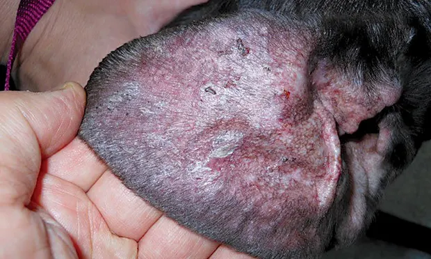
Figure 1A.
At presentation, there was a generalized and diffuse loss of hair coat with fine scaling (A). Thicker crusts were noted on the dorsum of the body (B). The pinnae were erythematous, with patches of adherent scale and crust (C).
Diagnostic Results
Samples for routine blood work and histopathologic examination of skin biopsies were submitted for this patient.
CBC and serum chemistry panels were normal except for an elevated globulin of 5.1 g/dL (reference range [RR], 2.4-4.0), and total protein of 7.8 g/dL (RR, 5.5-7.5), and mild thrombocytopenia of 91 × 103/µL (RR, 143-448 × 103/µL).
Histopathologic abnormalities from skin tissue included moderate acanthosis with laminated-to-compact hyperkeratosis and a superficial dermal mild-to-moderate band-like infiltrate of lymphocytes, pigmented macrophages, plasma cells, and few neutrophils. Sebaceous glands were absent. There was blurring of the dermoepidermal junction, and the basal cell layer showed extensive squamatization with mild vacuolar alterations and scattered apoptotic keratinocytes.
Ask Yourself
Based on the clinical findings and history, which of the following differential diagnoses is the least likely?
Staphylococcal folliculitis (pyoderma)
Discoid lupus erythematosus
Cutaneous leishmaniasis
Exfoliative cutaneous lupus erythematosus
What are the most appropriate diagnostic test(s) to consider in this case?
Skin scraping for cytologic examination
Routine CBC and serum chemistry panel
Skin biopsy for histopathologic examination
All of the above
Pending test results, which treatment option would be the most appropriate to begin?
Cephalexin 25 mg/kg twice a day
Ketoconazole 10 mg/kg once a day
Prednisolone 0.5 mg/kg twice a day
Chlorhexidine-containing shampoo every other day
Did You Answer?
The least likely differential diagnosis is discoid lupus erythematosus, an autoimmune disease characterized by depigmentation and crusting affecting the nasal planum, lip margins, and eyelid margins. Crusts often extend onto the pinnae and dorsal muzzle. Lesions are primarily confined to the head, and systemic signs are absent; in this case, lesions were generalized, and the nasal planum was not affected. Clinical lesions in this case are similar to those found in cutaneous leishmaniasis. Although not a disease endemic in the patient’s current location, the travel history before adoption was unknown. Staphylococcal folliculitis (pyoderma) typically presents with pustules progressing to crusted papules. Over time, lesions enlarge to produce punctate alopecic patches with peripheral scale and epidermal collarettes. This patient’s lesions consisted of generalized and diffuse fine scaling with no evidence of epidermal collarettes. Diffuse scaling can develop with chronic superficial pyoderma, especially in cases concurrently treated with corticosteroids, but there was no reported response to antiseptic shampoo therapy or to antibiotics.
All of the diagnostic procedures listed are appropriate in this case. Demodicosis can cause patches of alopecia with inflammation and crusts. Blood work is appropriate in order to establish a baseline prior to treatment as well as to assess potential causes of systemic signs in this case. The benefit of skin biopsy here is to establish a definitive diagnosis and/or to exclude other differential diagnoses. Skin biopsies are often obtained when in-house diagnostics are negative and initial therapy is not effective.
A chlorhexidine-containing shampoo every other day is the most appropriate first treatment option. Without confirmation of either a bacterial or a fungal skin infection, empiric treatment with systemic antiinfection medications is questionable. Conversely, treatment with prednisolone before the exclusion of infection would be detrimental in this patient as corticosteroids may worsen ongoing infection and/or occult demodicosis.
Diagnosis
Exfoliative cutaneous lupus erythematosus (ECLE)
Discussion
ECLE is a familial form of lupus found in German shorthaired pointer dogs. ECLE is a unique, heritable disease that causes signs similar to subacute cutaneous lupus erythematosus in human beings.1 Lesions develop in young adult dogs (ages 6 months to 2 years 9 months) consisting of scaling and alopecia initially affecting the face, pinnae, and dorsum with eventual generalization. In addition, dogs often exhibit lameness, pain, and pyrexia. Anemia and thrombocytopenia develop in chronic and severe cases.2
An autosomal recessive mode of inheritance caused by a mutant gene on canine chromosome 18 has been proposed.1 IgG deposition at the basement membrane and circulating antifollicular and anti-sebaceous IgG antibodies have also been identified.3
The prognosis for affected dogs is poor-to-guarded.4 Treatments include topical antiseborrheics, essential fatty acid supplements, tetracycline–niacinamide, hydroxychloroquine, cyclosporine, and adalimumab, with poor overall success.4-7 Transient response to corticosteroids with azathioprine was reported in 1 dog.3 Most patients are euthanized by 4 years of age because of progressive dermatitis and worsening joint pain and stiffness.6
A previous patient diagnosed with ECLE has been successfully treated with leflunomide by the author for over 4 years. Because of a reported poor prognosis and the lack of response to previous therapy, treatment with leflunomide was initiated in this patient. Leflunomide has antiproliferative activity for rapidly reproducing cells—especially lymphocytes—inhibiting autoantibody production by B-cells and decreasing proliferation of autoimmune T-cells.8
Related Article: Canine Leishmaniasis: An Overview
Treatment & Outcome
Initial leflunomide treatment dosage was 80 mg/day (3.5 mg/kg/day). At 4 weeks, lesions were significantly less erythematous, and the patient was reported to be less painful and less depressed. However, significant pruritus developed and the patient was prescribed oclacitinib 9.8 mg twice a day (0.5 mg/kg twice a day) for 14 days, reduced to once a day for maintenance of comfort. After 1 year of treatment, the patient remains stable and comfortable, with no signs of malaise or stiffness. The patient continues to be administered leflunomide 80 mg every other day and oclacitinib 9.8 mg daily (Figure 2). To the author’s knowledge, this article is the first to report the successful treatment of ECLE with leflunomide. Additional case experience using leflunomide, with or without oclacitinib, is needed to document the efficacy of this treatment for ECLE.

Figure 2A.
After 1 year of treatment with leflunomide, the hair coat appears normal and shiny (A), with near-normal hair coat regrowth on the dorsum (B). The pinnae appear normal, with hair coat regrowth on the concave surfaces and with an absence of scales (C).