Changes in Feline Eating Behavior
Case 1History. A 5-year-old, neutered male domestic shorthair that lives indoors with 2 other cats was presented for decreased willingness to eat dry food. When he did eat dry food, he would often drop the kibble. No previous dental procedures had been done. Wet food was well tolerated.
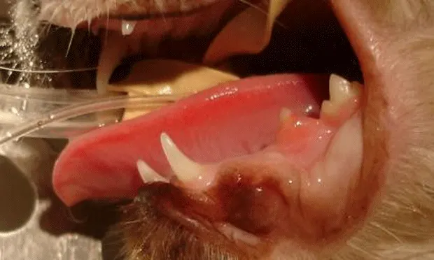
Physical Examination. General physical examination was normal. Oral examination revealed gingival hyperplasia around lower right third premolar (407) and partial crown of lower right first molar (409) (Figure 1, above). Touching either of the 2 teeth with a fingernail elicited a pain reflex.
Case 2**History.** A 4-year-old, spayed female ragdoll that lives indoors and is the only pet was presented for a change in eating behavior. Despite a long-standing preference for dry food, the cat now would eat only wet food. The owners had noticed some shyness when they attempted to stroke the cat's face and forehead.

Physical Examination. General physical examination was normal. There was mild calculus on both upper fourth premolars (108, 208) and gingival hyperplasia with gingival edema of the lower left third premolar (307) (Figure 2, right, View larger image). The lower right third premolar (407) was missing. Pain was elicited when 307 was tapped lightly with a fingernail.
ASK YOURSELF ...• Why are these cats reluctant to eat dry food?• Why are pain reflexes elicited?• What differential diagnosis is consistent with the clinical signs and oral examination?• What diagnostic tests should be done?• What should be the treatment plan?
Diagnosis: Feline tooth resorption
Diagnostics.For both cats, all preoperative CBCs and blood chemistry indicators were within normal limits. Full-mouth intraoral radiographs were taken during general anesthesia.
Related Article: Feline Tooth Resorption—Extraction of Retained Premolar Root

Case 1: Roots of teeth 407 and 409 show evidence of root ankylosis and replacement resorption. A discernable periodontal ligament space is absent. Crowns are affected (Figure 3, left View larger image).

Case 2: Tooth 307 shows loss of crown substance and partial resorption of distal root. Mesial root has a discernable periodontal ligament space and partially discernable periodontal ligament space around portions of the distal root (Figure 4, right View larger image).
Related Article: Digital Dental Radiology
Differential Diagnosis. Gingivostomatitis is a very common and painful oral condition in cats, but the lack of inflammation of the mucosal tissue near the upper and lower teeth or in the caudal part of the mouth decreases the likelihood of this condition. Also, with gingivostomatitis, pain is normally present when opening the cat's mouth, not just when tapping on a suspected tooth. Acute pain on tapping the tooth and lack of attachment loss of gingival and bony tissues around the teeth make periodontal disease a less likely diagnosis.
Treatment**Case 1:** The crowns of both 407 and 409 should be amputated to the level of the alveolar ridge and the roots left for continued replacement resorption.
Case 2:Tooth 307 should be removed completely, leaving no roots behind.
It has often been stated that crown removal is all that is required when treating feline tooth resorption. Unfortunately, this is not always the case because, as illustrated by these patients, there are 2 types of clinical presentations of tooth resorption in cats. In the first type (Case 1), affected roots are incorporated into the process of bone remodeling, leading to gradual resorption and replacement of tooth substance by bone (replacement resorption). The second (Case 2) exhibits root resorption characterized by simultaneous resorption of alveolar bone adjacent to the tooth as well as a radiographically visible periodontal space around unaffected roots.
Proper treatment of tooth resorption is determined with the aid of intraoral radiographs to ascertain the type of resorption. With this all-important diagnostic aid, complete extraction or crown amputation of affected teeth can be chosen appropriately.
OutcomeBoth cats returned to normal eating and activity.
DID YOU ANSWER ...• Oral pain• Exposure of sensitive tooth structures (dentin, cementum, and/or pulp)• Feline tooth resorption• Intraoral radiographs• Case 1: crown amputation; Case 2: complete extraction of tooth
CHANGES IN FELINE EATING BEHAVIOR • Johnathon Robert Dodd
Suggested Reading
Acquired defects: Caries and regressive changes. Mulligan TW, Williams CA, Aller MS. In Aller MS (ed): Atlas of Canine & Feline Dental Radiography-Trenton, NJ: Veterinary Learning Systems, 1998, pp 160-169.Crown amputation with intentional root retention for advanced feline resorptive lesions-A clinical study. DuPont G. J Vet Dent 12:9-13, 1995.Crown amputation with intentional root retention of dental resorptive lesions in cats. DuPont G. J Vet Dent 19:107-110, 2002.Feline oral and dental disease. Lobprise HB, Wiggs RB. Veterinarians Companion for Common Dental Procedures-Lakewood, CO: AAHA Press, 2000, pp 140-146.Prevalence of odontoclastic resorption lesions and periapical radiographic lucencies in cats: 265 cases (1995-1998). Lommer M. JAVMA 217:1866-1869, 2000.Update on the etiology of tooth resorption in domestic cats. Holmstrom SE. Vet Clin Small Anim 35:913-942, 2005.