Central Venous Catheter Placement: Modified Seldinger Technique
A central venous catheter (CVC) is a large diameter catheter that can be placed in the jugular or peripheral vein. CVCs can be indicated for various procedures, including central venous pressure monitoring, blood sampling, parenteral nutrition, and IV fluid and hypertonic solution administration.<sup4sup>
The following focuses on CVC placement using the modified Seldinger technique, a common indication in veterinary patients.
The original Seldinger technique was developed by radiologist Sven Ivar Seldinger in 1953 to allow large-bore catheter placement in peripheral arteries for angiography. Before the Seldinger technique, this procedure required a large-bore needle (through which a narrower bore catheter was threaded), restricting its use to larger arteries and making vessel puncture more difficult.1
Original Seldinger Technique
The Seldinger technique’s addition of a flexible, round-ended, metal leader (guidewire) was unique.1 The sequence included needle puncture, guidewire threaded through needle, needle removed, flexible catheter threaded over guidewire, and guidewire removed.1 This process enabled a catheter of the same bore as the needle to be inserted percutaneously, rather than requiring surgical exposure or a large bore needle.
The original Seldinger technique has been associated with several complications including inadvertent catheter placement into the internal thoracic vein, azygous vein, and caudal vena cava.2 Perforation of vessels and/or the right atrium has also been reported.2 Patency of the central venous system should not be considered sufficient evidence of adequate placement.2 Radiography should always be performed to confirm appropriate placement.
Modified Seldinger Technique
The modified Seldinger technique allows for placement of large-bore catheters in jugular veins, the pleural space, pulmonary arteries, and even hollow organs. It is also often used to place dialysis catheters. The primary modification was to use an introducer catheter (rather than a needle) to minimize entering other vessels. Other modifications have included the use of peel-away catheters and variation in stylet, catheter, and wire materials and lengths. Placement of CVCs using the modified Seldinger technique is commonly performed in veterinary patients.
Complications & Considerations
Complications associated with CVCs include embolism, thrombosis, infection, migration or loss of the guidewire, and mechanical complications (eg, kinking, catheter fracture, catheter removal by the patient). The most common types of bacteria associated with catheter infections are Streptococcus spp, Staphylococcus spp, Escherichia coli, and Enterobacter spp.3 Appropriate sterility (ie, surgical scrub, drapes, cap, mask, gloves) must be maintained. Wearing gloves during catheter manipulation and keeping the bandage around the site clean and dry are appropriate measures to decrease risk for catheter-associated infections.
Step-by-Step: Placing a Central Venous Catheter Using the Modified Seldinger Technique
What You Will Need
Catheter placement kit that includes:
18-gauge, 1.5-inch catheter (of appropriate size for patient)
Multilumen or single lumen over-the-wire catheter (of appropriate size and length for patient)
Guidewire
Vessel dilator
Scalpel blade (#10 or #11)
6-mL syringe
Suture wings and suture wing clips
Chlorhexidine 2% scrub
Alcohol
Sterile gloves and drapes
Nonabsorbable suture (2-0 or 3-0 nylon)
Bandage material
Syringes with heparin saline flush
Scissors, needle driver, and thumb forceps
Clippers


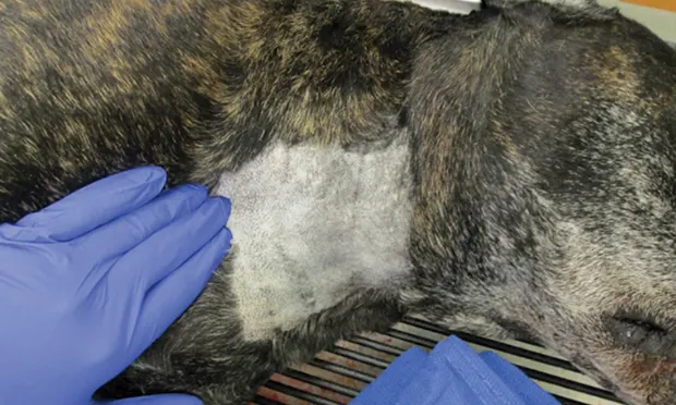
STEP 1A.
Place the patient in lateral or dorsal recumbency and identify the jugular vein. Mild to moderate sedation is often sufficient, although some patients may require general anesthesia. Appropriate monitoring is always required. Arrhythmias are possible, as the guidewire and/or catheter may be inadvertently advanced into the heart, making electrocardiogram monitoring essential. Prepare the site aseptically using surgical scrub technique with 2% chlorhexidine scrub alternating with alcohol.