The Case: Persistent Wound in a Cat
History
First PresentationA 5.3-kilogram, 3-year-old spayed female domestic shorthair cat is presented for wound on ventral abdomen, possibly from a bite. Cat tests negative for feline leukemia virus/feline immunodeficiency virus.
Abdominal radiographs: Unremarkable, no evidence of abdominal involvement
Wound explored/flushed; drain placed
Treatment 1: Amoxicillin/clavulanic acid (62.5 mg q12h PO) × 7 days
Drain removed 3 days later
One week later, wound not healing well
Treatment 2: Marbofloxacin (25 mg q24h PO) × 10 days
The wound heals uneventfully.
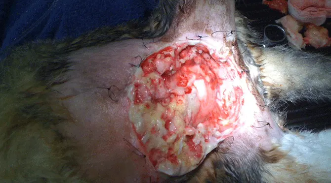
Figure 1 (above). The wound at culmination of the slough. Sutures are in place around the borders of the lesion in preparation for wet-to-dry bandages.
Second PresentationCat develops a draining tract in the same area 16 months later.
Temperature: 103.1⁰F
Treatment: Marbofloxacin (25 mg q24h PO) × 10 days along with warm compresses
The draining tract appears to resolve.
Third PresentationAgain, 7 months later, the cat develops for swelling in the area.
Temperature: 102.2⁰F
Treatment: Surgery to debride area is discussed with owner but declined.
Cefovecin sodium (40 mg SC)
Marbofloxacin (25 mg q24h PO) × 10 days
The swelling resolves.
Fourth PresentationEight months later (now nearly 3 years after initial presentation) the cat is presented with ventral abdominal swelling once again.
Temperature: 103.5⁰F
CBC/serum biochemistry profile/UA: Unremarkable
Treatment:Excisional surgery of the affected area is recommended and performed the following day. The affected area is excised down to the body wall and submitted for biopsy, and a sample for culture is taken from the center of the affected area.
Drain placed/cefovecin sodium (40 mg SC) administered
Drain removed 3 days later
Marbofloxacin (25 mg q24h) × 10 days initiated
Two days after drain is pulled, cat is presented for swelling in the area/dehiscence of surgical closure. Over next day or 2 day, the entire area of the ventral abdominal fat pad becomes necrotic and sloughs.
Biopsy results: Chronic suppurative steatitis/panniculitis/cellulitis
Anaerobic/aerobic culture: Negative

Treatment
Area completely debrided/anchor sutures placed to hold a wet-to-dry bandage in place
Doxycycline (50 mg q24h PO)/marbofloxacin (25 mg q24h PO) initiated
Daily wet-to-dry bandage changes until area is ready for closure (14 days)
Figure 2. After wound closure
Recovery/Outcome
Final wound closure achieved 15 days after wet-to-dry bandages initially applied
Doxycycline/marbofloxacin continued for another 30 days
No recurrence after 14 months
The Specialist's Opinion
Assuming that this was a bite wound, there is a high likelihood that it was more severe than was suspected based on external appearance. The twisting and shaking motion delivered in a bite can produce extensive damage, shear direct cutaneous vessels, and inoculate bacteria deep within the tissues. In this case the veterinarian started out with some excellent diagnostic tests: evaluation for viral diseases that could result in immunosuppression and assessment of the abdominal cavity for evidence of internal damage. It may have been useful to evaluate the cat for systemic illness to predict problems with wound healing and determine whether additional care such as fluids or oncotic support was warranted.
Initial wound management is not described in detail, so it’s difficult to assess whether it was sufficient. Use of amoxicillin/clavulanic acid was reasonable, since the most common organisms found in bite wounds are initially sensitive to this agent. Once a bite wound becomes nonhealing or recurrent, however, a more aggressive approach is required. The trick is to convince owners that a little more investment of time and money now can prevent the need for serious intervention in the future.
Bite Wound Management
Bite wounds should initially be managed by clipping, prepping, and draping the area as for aseptic surgery. If puncture wounds are small, a sharp incision is made through them to allow insertion of a sterile gloved finger or instrument to probe the depths of the wound and evaluate the extent of trauma. Puncture wounds should not be flushed unless they can be opened: infusion of saline through small wounds will cause dissemination of bacteria and fluid into deeper tissue planes.
Once the wound is cleaned and flushed, a tissue sample should be obtained for culture. The veterinarian must then decide on primary closure or open wound management. Small wounds with minimal tissue damage and debris can be left open to heal. Wounds with extensive tissue damage require debridement. If foreign material and damaged tissue can be fully removed, wounds can be closed over a drain. If not, they are managed open until they heal on their own or are healthy enough for closure. Penrose drains must be placed through a fresh skin incision in the most dependent portion of the region, and the drain exit site must be covered with a sterile wrap that is changed at least every 24 hours.If drains are not covered, ascending infection will occur, and if drains are not dependent, fluid accumulation will delay healing and potentiate infection. While more expensive, continuous suction drains provide a closed system that can be inserted in a nondependent region, facilitating bandage placement.
Nonhealing Wounds
If a wound fails to heal or reoccurs, the veterinarian must assess for primary or predisposing causes of infection or inflammation. Cats that are negative for FeLV or FIV on one test may later become positive. Those that have systemic disease such as nutritional deficiencies; anemia; or hepatic, renal, or cardiac dysfunction are more likely to have difficulty with wound healing. If a fistula or sinus tract is present, a radiographic or CT contrast study may help determine the source of the drainage.
Once diagnostics are complete, the wound should be surgically explored and debrided and the tissues sampled. A deep tissue culture is critical for ruling out resistant infection and more unusual microbes such as Mycoplasma, Mycobacterium, and Bartonella species; Coccidioides immitis; and Sporothrix schenckii. Detection of these organisms may require multiple cultures. Tissue can also be submitted for cytology and biopsy to look for unusual organisms, foreign material, and evidence of ischemia or extensive inflammation.
Secondary Panniculitis & Other Complications
Cats can develop chronic, nonhealing wounds secondary to sterile panniculitis, as was noted in this patient, or entrapment of hair and other debris. Even with removal of the reactive tissues and improvement of the blood supply by omentalization, these wounds may dehisce with primary closure. Therefore, as in this cat, open wound management may be the best treatment. Use of vacuum-assisted closure or topical dressings such as honey will speed the formation of granulation tissue, which responds well to moisture. Many clinicians use wet-to-wet (or, more appropriately, moist-to-moist) bandages for this reason. Systemic antimicrobials are necessary when cellulitis secondary to infection arises. Since this cat was on antibiotics at the time of the culture, it is impossible to know whether systemic antibiotics were necessary.
One important question about this patient is its rabies vaccination status in the face of the probable bite. If the cat was unvaccinated or its vaccines were overdue at time of the trauma, vaccination and quarantine would be recommended, with the duration of isolation based on state law (usually 6 months). Cats that are current with their rabies vaccines should be revaccinated and quarantined for 45 days.
Karen Tobias, DVM, MS, DACVS, is a referral surgeon and professor of surgery at University of Tennessee Veterinary Medical Center. Dr. Tobias also taught at University of Georgia and Washington State University. She is the author of Manual of Small Animal Soft Tissue Surgery and coeditor of Veterinary Surgery: Small Animal. She graduated from University of Illinois College of Veterinary Medicine and completed an internship and residency at Purdue University and Ohio State University, respectively.