The Case: Mysterious Disappearing Foreign Body

History
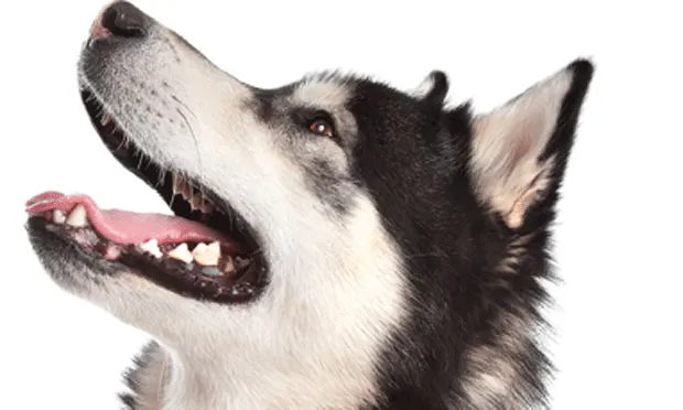
A 2-year-old intact male Malamute with a history of consuming foreign objects. Half a ball had been removed from its stomach 4 months earlier.
Presentation
Patient was presented with vomiting/inappetence/lethargy of 2 days’ duration. Client voiced financial concerns. (8/14)
Abdominal radiographs revealed distended stomach filled with homogeneous material/no further foreign material identified. Client asserted patient had not had anything by mouth for 2 days.
Fecal testing from initial presentation 4 months previously: +1 Clostridium, +3 spirochetes/motile bacteria. No fecal test done on this presentation.
Exploratory laparotomy revealed stomach full of water, which was removed by stomach tube. Client then remembered dog had drunk a bowl of water prior to exam. Palpation of length of GI tract from stomach through exteriorized intestines (pylorus to ileocecal junction) repeated several times. Nothing found.
In face of financial constraints, a sample for histopathology was not obtained.
Recovery
Patient recovered from surgery with no appetite. Had soft scant stools/improved spirits.
Postsurgical Treatment
Fluid therapy before/during/after surgery: lactated Ringer’s solution
8/14:
Penicillin (300,000 U/mL): 5.5 mL SC
Gentamicin (100 mg/mL): 0.5 mL SC
Maropitant citrate (10 mg/mL): 3.0 mL SC
8/15–8/18:
Sucralfate: 1 g q12h PO
Maropitant citrate: 60 mg q24h PO
Metronidazole: 500 mg q12h PO
Amoxicillin: 500 mg q12h PO
8/16:
Patient ate on its own (full can i/d and a/d mixed)
8/17:
Stopped eating/lethargic
Force fed half can a/d
Mirtazapine: 15 mg PO (one time dose)
8/18:
Still not eating
Continued oral meds (above)
Radiograph: distended stomach, food passing through bowel
Sedated to pass stomach tube: Removed gas/fluid; radiograph afterward showed normal stomach
SNAP cPL Test (canine pancreas-specific lipase): Negative
CBC/serum analysis: WNL
IV fluids: lactated Ringer’s solution @ 50 mL/hr started; decreased to 30 mL/hr ~ 90 min later
Prednisolone injectable: 100 mg IV slow
Discontinued amoxicillin
NPO in evening
Treatment8/19–8/24:
Famotidine: 20 mg q12h PO X 2 days; q24h X 2 days; q12h for 2 days
Metronidazole: 500 mg q12h PO X 3 days; q24h X 1 day; then q12h for 1 day (discontinued evening dose on 8/24)
Sucralfate: 1 g q12h PO X 3 days; then q24h X 1 day; then q12h X 2 days)
IV fluids
Lactated Ringer’s solution with 3.0 mL (100 mg/mL) vitamin B complex at varying rates X 2 days
Continued lactated Ringer’s solution @ 75 mL/hr X 3 days
Maropitant citrate reinitiated (8/20): 60 mg q24h PO; none X 1 day; q24h X 2 days; then discontinued
Penicillin (300,000 U/mL): 5 mL SC (8/19)
Gentamicin (10 mg/mL): 0.5 mL SC (8/19)
Vitamin B12 (100 mg/mL): 0.6 mL SC (8/20)
Metoclopramide (5 mg/mL): 0.8 mL SC initiated 8/23; then q12h X 1 day
Mirtazapine: 15 mg PO (8/22–8/24)
Nutrition: Force fed canned a/d (8/21–8/22); offered canned a/d (little interest) 8/23 and a/d dry & canned (8/24)—ate 1 cup dry
Removed IV catheter 8/23; offered small amount of water
Monitoring
Bowel Activity: None AM despite posturing, small volume PM on 8/19; scant soft feces 8/20; none 8/21 & 8/22; scant diarrhea 8/23–8/24
Urinated daily in varying amounts 8/19-8/24; ate ice cubes 8/24
Diagnostics
Barium radiograph (8/23) after 4 daily doses barium (20 mL PO)
Serum biochemical analysis (8/18): Normal
CBC (8/18): Normal
Activity
Client took dog with her for a few hours to get it out of clinic (8/24). Closely watched during outing.
8/25:
Urinated only in AM, wanted to eat grass
Sucralfate: 1 g PO
Famotidine: 20 mg PO (AM only)
Offered i/d canned and dry in separate bowls, no immediate interest. Found both bowls empty about an hour later.
Offered ice cubes; ate readily
Metoclopramide (5 mg/mL): 0.8 mL q12h SC
Rectal temperature: 103.2⁰F (normal until today)
Discharged with chart and radiographs to another veterinary clinic for second opinion. Diagnosed as oxygen deprived, also given Lactobacillus acidophilus
Transferred to third veterinarian in another city
Outcome
Third veterinarian contacted our clinic: Patient had developed a fever, thus they performed repeat surgery 25 days after initial presentation at our clinic. A piece of a tennis ball was found in the small intestines along with signs that a perforation had occurred and later healed proximal to the obstruction; 9 inches of bowel was removed.
Patient recovered with a healthy appetite.
The Specialist’s OpinionEric Pope, DVM, DACVS
Given the history and the owner’s financial concerns, it would be easy to assume this onset of vomiting was likely due to foreign body ingestion. One could argue that with the relatively short duration of clinical signs and the fact that abdominal radiographs showed a distended stomach with no evidence of obstruction, conservative management could have been instituted and the response to treatment assessed. The downside of this approach is that the delay would allow more time for a foreign body to move from the stomach into the intestine.
Financial ConcernsFinancial concerns are often an issue but can’t overshadow best-care practices and procedures. In fact, the cost of treatment can end up being higher if indicated diagnostic measures are omitted, as probably was the case with this patient. Preoperative endoscopic and/or ultrasonographic examination could have provided very useful information. Since there was no evidence of intestinal obstruction, my preference would have been to recommend or perform endoscopy to minimize costs. There is a high likelihood that the foreign body was still in the stomach at this time and that it could have been retrieved noninvasively. If no foreign body was present, endoscopic biopsy of the stomach and proximal intestine could have been performed.
Celiotomy & GastrotomyIf performing other diagnostics was not an option and the owner declined conservative management, exploratory celiotomy would be the next step. Identification of nonlinear foreign bodies in the intestine by visual appearance and palpation is probably pretty sensitive, and repeating the examination multiple times reduces the likelihood a foreign body(ies) will be missed.
However, evaluation of the stomach by palpation alone is probably more likely to result in failure to identify a foreign body, especially if the item is small or composed of soft material. The presence of other contents in the stomach, as in this case, also makes identification of a foreign body by palpation alone more difficult. The size of the piece of tennis ball ultimately removed was not described, but, if it was small enough to exit the stomach, it may have been difficult to palpate through the stomach wall.
In a properly prepared patient, the risk of complications following gastrotomy is low and this operation would have been indicated to completely explore the stomach. If the index of suspicion for a foreign body was sufficiently high to warrant surgical exploration, there was justification to perform a gastrotomy to explore the stomach thoroughly.
The Role of PainThe postoperative pain management protocol was not described: pain can influence gastric function and could account for the stomach distention postoperatively. The gastric paresis and distention could result in nausea and affect the patient’s appetite.
Eric R. Pope, DVM, MS, DACVS, is professor of Small Animal Surgery and section head of Small Animal Clinical Sciences at Ross University School of Veterinary Medicine. A general surgeon, Dr. Pope focuses his research interest on wound management and reconstructive surgery, surgical oncology, and minimally invasive techniques for thoracic and abdominal surgery. He has authored or coauthored 50 publications in referred journals and numerous book chapters, serves on the editorial review board of several journals, and speaks widely at international and national meetings. Having earned his DVM from Auburn University, Dr. Pope joined a small animal practice in Jacksonville, Florida; then returned to Auburn University to complete a surgery residency and Master of Science degree program. He served on the faculty of the University of Tennessee and University of Missouri before joining the staff at Ross University in 2007.
The Generalist’s OpinionBarak Benaryeh, DVM, DABVP
It’s important during any exploratory to take every precaution not to miss a foreign body. The stomach should be carefully palpated and bowel checked from the duodenum to the colon. The two trickiest areas are near the duodenocolic ligament and at the cardia within the stomach. Foreign bodies high in the stomach can be very difficult to palpate or can slide up into the esophagus during surgery. Some veterinarians espouse a gastrotomy during any procedure where there is a high suspicion of a foreign object. Though extreme, this procedure ensures the stomach can be completely checked.
Concentrating on the Postoperative PeriodThe focus of this case should not be on the initial procedure: this dog could have eaten the tennis ball fragment postoperatively or the fragment could have been within the esophagus. The learning points are in the days following the surgery and determining what treatments and diagnostics are best performed when a surgical patient fails to recover as expected.
Two days after surgery, this dog did eat on its own, which likely led to a delay in further diagnostics. Whether or not a foreign body was present during the initial procedure, it is clear this dog’s recovery did not go as planned. When the dog was still inappetent 4 days after surgery, 100 mg of injectable prednisolone was administered. Injectable steroids postoperatively could slow the healing process and put the patient at risk for further gastrointestinal complications.
Also at 4 days, postoperative plain radiographs were taken and a SNAP cPL performed. Although these tests were negative in this case, one should note they can be misleading. Foreign bodies (especially in the duodenum) can lead to secondary pancreatitis and a positive cPL test can precipitate a focus on the pancreas rather than the foreign body. These tests should not be relied on for a definitive diagnosis.
ImagingNine days after the initial procedure, barium was given in 4 doses of 20 mL each. Opinions differ on a diagnostic dose of barium, varying from 5 mL/kg to 3 to 6 mL/lb, but even with conservative dose estimation, 80 mL would not be enough barium for a dog this size. In addition the barium should be given in a single dose rather than 4 separate increments. If there is any risk of a perforation, barium is contraindicated.
If available, an ultrasound is generally less costly and easier to perform than a barium series and can be highly diagnostic for gastrointestinal foreign bodies. Ultrasound is, however, highly operator-dependent, so ideally it should be performed by a board-certified radiologist.
The fact that 25 days later there was evidence of a healed perforation does not necessarily relate to this presentation. The perforation could have occurred at any time, such as when the first foreign body was removed 4 months prior to the inciting cause of this presentation. It seems unlikely that this dog experienced a perforation and remained stable.
Take-Home LessonsFortunately the patient did pull through. There is no way to know what the perfect way to have handled this case would have been; the take-home lessons are to have a methodology for exploratories and a comfort level with how best to handle postoperative complications.
Barak Benaryeh, DVM, DABVP, is the owner of Spicewood Springs Animal Hospital. He graduated from University of California–Davis School of Veterinary Medicine in 1997 and completed an internship in Small Animal Medicine, Surgery, and Emergency at University of Pennsylvania. Dr. Benaryeh has also taught practical coursework to first-year veterinary students and was a primary veterinary surgeon for the Helping Hands Program, which trains assistance monkeys for quadriplegic people. Dr. Benaryeh is certified by the American Board of Veterinary Practitioners in Canine and Feline Practice.