A Case of Cough, Lethargy and a Tracheal Wash
A 4-year-old, female spayed Labrador retriever mix was presented for cough, lethargy, and decreased appetite of 1-week duration.
Physical examination showed depression, weight loss, and harsh lungs sounds. A moderate, diffuse, nodular interstitial pattern was identified on thoracic radiographs. Fluid collected from a tracheal wash was submitted to the diagnostic laboratory for cytologic evaluation. Concentrated smears were examined (Figures 1 to 3).
ASK YOURSELF ...
• What are the expected cytologic findings in tracheal fluid from a normal dog?• What are the differential diagnoses?• What are the distinguishing cytologic features of the etiologic agent?
Diagnosis: Blastomycosis
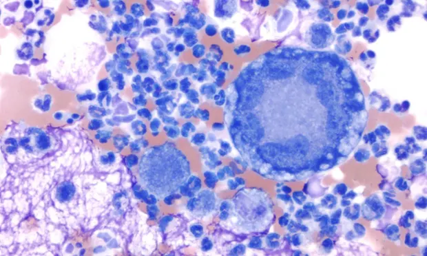
Cytologic evaluation. Smears were highly cellular and contained many inflammatory cells (Figure 1), moderate numbers of erythrocytes, and occasional clusters of columnar epithelial cells (Figure 2) in a thick background of mucus. The inflammatory cells were primarily neutrophils with moderate numbers of macrophages and relatively few multinucleated giant cells, lymphocytes, and plasma cells.
The epithelial cells were well-differentiated. Occasional extracellular yeast organisms (Figure 3) were identified. These organisms were round and 10 to 20 µm in diameter with a thick, blue wall and granular blue internal contents. Broad-based budding was common. No other etiologic agents were observed.
Diagnosis. Definitive diagnosis is made by cytologic or histologic identification of the organism.1,2 Infection typically causes a suppurative to pyogranulomatous inflammatory response.3 Choice of tissue sample depends on clinical signs and affected sites. In dogs with respiratory signs, tracheal wash or bronchoalveolar lavage may be useful; however, most samples are not diagnostic due to the interstitial location of the organism.4 If either procedure is negative, fine-needle aspiration of the lungs may reveal organisms. Fine-needle aspiration or impression smears of affected lymph nodes, cutaneous or subcutaneous lesions, bone, or vitreous are diagnostic in most dogs.2,4
Serologic testing should be done if organisms have not been identified after repeated cytologic or histologic examination of affected tissue.1,2 Titers may be negative during the early phase of infection. Positive titers may persist after successful resolution of infection. Thoracic radiographic findings often include diffuse to nodular interstitial, bronchointerstitial, or alveolar patterns in the lung and tracheobronchial lymphadenopathy. In dogs with bone involvement, radiographs reveal osteolysis, periosteal proliferation, and soft tissue swelling. Routine blood test results are nonspecific. CBC findings may include mild nonregenerative anemia and neutrophilic leukocytosis with or without a left shift. The most common abnormalities on a biochemical profile are hypoalbuminemia and hyperglobulinemia. Hypercalcemia is present in a small percentage of dogs.1,2,4
Foundation of Blastomycosis
Pathophysiology: Systemic disease in dogs caused by the dimorphic fungus Blastomyces dermatitidis; infection usually occurs by inhaling mycelial spores1; spores are transformed into the yeast phase after phagocytosis by alveolar macrophages; organism may remain localized in lung or disseminate systemically via blood or lymphaticsCommonly affected distant sites: Skin, eyes, bones, lymph nodes, and subcutaneous tissue1,2
Clinical signs: Often nonspecific, including weight loss, lethargy, anorexia, depression, and fever; respiratory signs are common and include cough, tachypnea, dyspnea, and harsh lung sounds on auscultation2,4
Did You Answer ...
• Tracheal washes from healthy dogs typically have low cellularity and contain primarily ciliated and nonciliated columnar to cuboidal epithelial cells, macrophages, and a small amount of mucus. Neutrophils usually comprise less than 5% of the nucleated cell population. Other cells found in low numbers include lymphocytes (< 15%), eosinophils (< 5%), and mast cells (< 2%).3 Direct and concentrated smears of the fluid should be made as soon as possible after collection because cells deteriorate in vitro.• Pyogranulomatous inflammation occurs with fungal infections (e.g., blastomycosis, coccidioidomycosis, aspergillosis, histoplasmosis), protozoal infections, or foreign body reactions.3 Mixed inflammatory reactions are also found with necrosis associated with neoplasia or lung torsion.• The distinguishing cytologic features of the yeast form of Blastomyces dermatitidis are the round shape; size (generally 5 to 20 µm); thick, biconcave wall; granular internal contents; and frequent broad-based budding.3 The organism stains a dark blue with Romanowsky-type stains (e.g., Wright's stain, Diff-Quik). Hyphal forms are rarely found in tissues.1
COUGH & LETHARGY • Jennifer S. Thomas
References1. Blastomycosis. Legendre AM. In Greene CE (ed): Infectious Diseases of the Dog and Cat, 2nd ed. Philadelphia: WB Saunders Co, 1998, pp 371-377.2. Update on canine and feline fungal diseases. Kerl ME. Vet Clin North Am Small Anim Pract 33:721-747, 2003.3. Respiratory tract. Burkhard MJ, Valenciano A, Barger A. In Raskin RE, Meyer DJ (eds): Atlas of Canine and Feline Cytology. Philadelphia: WB Saunders, 2001, pp 135-185.4. Blastomycosis in dogs: 115 cases (1980-1995). Arceneaux KA, Taboada J, Hosgood G. JAVMA 213: 658-664, 1998.