The Case: Acute Respiratory Distress
PRESENTATION 1
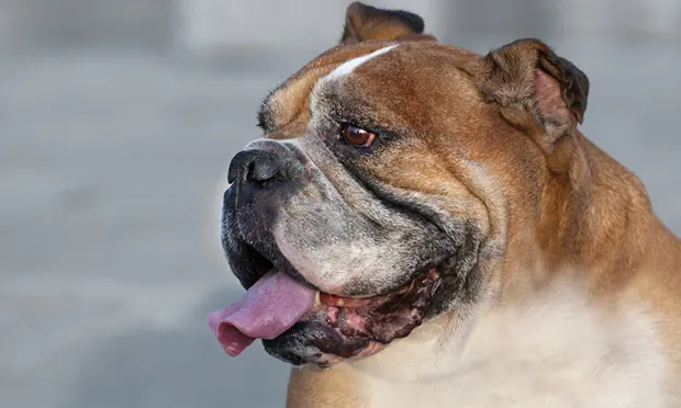
A 10-year-old intact male English bulldog presented for acute dyspnea (increased respiratory rate and effort) after becoming anxious at the sound of nearby fireworks. The patient had displayed intermittent panting over several days prior to this incident and had a 2-year history of hypothyroidism, currently controlled with levothyroxine 0.02 mg/kg PO q12h. He was current on vaccines and had no significant travel history.
Physical Examination
Bright, alert, but very anxious/nervous
Tachycardic (heart rate: 175 bpm)
Panting excessively, moderate stertor
Diagnostics
Pulse oximetry: 96%
Electrocardiogram: Sinus tachycardia (heart rate: 170–180 bpm)
Thoracic radiographs
Mild bronchointerstitial pattern, likely associated with age-related changes or chronic airway irritation
Heterogenous mineralization of the epiglottis, enlarged larynx/laryngitis, thickened soft palate, soft-tissue structure at dorsal aspect of the laryngopharynx. Differentials for these changes included edema/swelling or less likely an abscess or mass.
Sedated laryngeal exam: Mildly reduced to normal laryngeal function. Soft palate was elongated and a significant noise was heard as air rushed past. The soft palate changes were believed to be contributing to the narrowing of the functional upper airway but were not believed to be the primary cause for the acute crisis. There were also erythematous inflammatory lesions on the dorsal aspect of the epiglottis and along the arytenoid cartilages.
Treatment
Admitted for overnight monitoring
Butorphanol (0.2 mg/kg) administered IV when anxiety became progressively worse. Initially calmed but then became anxious again, leading to severe dyspnea and hyperthermia.
Anesthetic induction with propofol. Patient was intubated/ventilated until temperature returned to normal and he was breathing calmly under anesthesia.
At extubation, an antiinflammatory dose of dexamethasone (0.07 mg/kg IV) was administered; sedation was maintained with acepromazine (0.05 mg/kg IV) and butorphanol (0.2 mg/kg IV) until fully recovered from anesthesia.
Patient was discharged with tapering course of prednisone (0.25 mg/kg PO q12h x 5 days, then q24h x 5 days) and trazodone (1 mg/kg PO q12h) to manage noise-related anxiety.
Outcome
The patient had improved during hospitalization, so supportive outpatient treatment was chosen. The respiratory crisis was believed to have stemmed from a collection of factors including upper airway swelling/irritation; congenital, long-standing elongated soft palate; mild laryngeal dysfunction; and anxiety.
PRESENTATION 2
Patient presented 6 days after discharge for recurrence of dyspnea after the prednisone was tapered.
Physical Examination
No significant changes from previous exam
Diagnostics
Laryngoscopy: Elongated soft palate and thickened, edematous epiglottis
Abdominal ultrasound: Small splenic nodule, evidence of gastric ulceration/gastroenteritis, enlarged prostate, mass in left testicle
Treatment
Initially stabilized with acepromazine (0.05 mg/kg IM)
During anesthesia for laryngoscopy, soft palate was surgically trimmed with a laser. Biopsies were collected from the inflamed epiglottis with endoscopy.
Postoperative Treatment/Instructions
Cephalexin (20 mg/kg PO q12h)
Metronidazole (8 mg/kg PO q12h)
Prednisone (0.5 mg/kg PO q12h)
Discontinue trazodone
Outcome
Biopsy results: Focal predominantly fibrinous to mildly necrotizing sialadenitis/laryngitis with fibrous polypoidal hyperplasia. The main lesion was an area of prior distention and rupture of a minor salivary duct within the submucosa, initiating an area of edema, fibrin aggregation, minimal granulation tissue formation and mild necrosis, with overlying mucosal hyperplasia. There was no evidence of neoplasia or any infectious agents.
Prognosis: Good
At 3 months after discharge, the patient continued to do well on 0.5 mg/kg prednisone q12h. Decreasing the dosage of prednisone by 25% is currently under consideration.
The Specialist’s Opinion
Gretchen Statz, DVM, DACVECC
Initially this appeared to be a typical presentation of brachycephalic syndrome but turned out to have a unique diagnosis. In general, the case was handled well and thankfully the dog continued to thrive three months after discharge.
Brachycephalic SyndromeBrachycephalic syndrome is common in breeds (English bulldogs, Boston terriers, pugs) that display characteristic conformation and comprises a combination of anatomic abnormalities including elongated soft palate, stenotic nares, and hypoplastic trachea.1 These abnormalities lead to secondary complications including everted laryngeal saccules, laryngeal collapse, and everted tonsils.1 Brachycephalic syndrome most likely contributed to the respiratory distress in this case, but it was not the entire problem.
Differentials for LaryngitisInflammation of the larynx in the dog is not an uncommon finding and can have a variety of underlying causes, including:
Infection: Canine infectious tracheobronchitis or kennel cough
Trauma: External (bites, choke injuries) or internal (foreign bodies)
Endotracheal intubation
Insect bites
Neoplasia (relatively uncommon)
In many cases the cause of laryngeal inflammation is not obvious and the inflammation is self-limiting. This case was found to be initiated by a ruptured minor salivary gland. We were fortunate to have the benefit of a biopsy diagnosis. In a majority of cases, histopathology is not available. If more biopsies were performed in cases of laryngitis, it is possible that sialadenitis causing a local tissue reaction might be found to be more common.
Salivary Gland AnatomyIn all honesty, it was a surprise to me to learn (or perhaps remember) that there are salivary glands in the submucosa of the larynx. In review of basic anatomy, the salivary glands can be broken down into major and minor, with the major (parotid, mandibular, sublingual, and zygomatic) being those with which we are most familiar. There are also minor salivary glands throughout the oral cavity, including the submucosa of the larynx.
Emergency TreatmentDespite the underlying etiology, upper airway obstruction often requires aggressive intervention in the emergency setting. Sedation is extremely important to decrease both the panic caused by dyspnea and the respiratory rate/effort. Good sedatives can include a combination of benzodiazepines, opioids, and acepromazine, although the hypotension caused by acepromazine requires judicious use and close monitoring.
Hyperthermia is often an issue in patients with upper airway obstruction. Increasing effective ventilation, along with providing IV fluids or active cooling, as indicated, can help to cool the patient. If initial sedation fails to return the patient’s breathing to normal, induction and intubation are indicated. The duration of intubation can be anywhere from a few hours to overnight or longer––until the inflammation decreases or other interventions (surgery) can be performed.
Steroids can be helpful in situations of airway inflammation regardless of the underlying cause. A common mistake is to give an excessive dose of steroids but an inadequate dose of sedative drugs. The dose of dexamethasone used in this case was 0.07 mg/kg, which is equivalent to 0.5 mg/kg of prednisone (dexamethasone is 7 times more potent than prednisone; meaning 1 mg dexamethasone is equivalent to 7 mg prednisone). This is an appropriate antiinflammatory dose in this case.
A more aggressive sedative approach initially (adding a benzodiazepine or acepromazine) may have helped avoid the episode of severe dyspnea and hyperthermia that developed on the first night of hospitalization. Oxygen therapy is also indicated in cases of acute respiratory distress and was not mentioned in this case. Oxygen can be provided initially via flow by or mask and eventually through a nasal tube or nasotracheal tube (NTT). One study found that the use of NTT oxygen supplementation may minimize severe postoperative morbidity, in particular by reducing postoperative respiratory distress.<sup2 sup>
Long-Term TreatmentBecause this is a unique case, it is somewhat difficult to develop a long-term treatment protocol or predict the long-term prognosis. The underlying cause (rupture of the salivary duct) has likely resolved; therefore, weaning the prednisone is a good choice.
Gretchen Statz, DVM, DACVECC, is an internal medicine consultant for Antech Diagnostics and a VIN consultant on the internal medicine boards. A graduate of University of Wisconsin – Madison, Dr. Statz interned at VCA West Los Angeles and then worked for several years at two emergency/referral hospitals in the Boston area. After completing a residency at VCA Veterinary Referral Associates in Gaithersburg, Maryland, she became boarded in emergency and critical care. Having a strong interest in internal medicine, she has been practicing in that field for the past several years.
The Generalist’s Opinion
Barak Benaryeh, DVM, DABVP
An important consideration in discussing this case is a quick review of brachycephalic airway syndrome, which comprises a series of anatomic abnormalities. The conditions most commonly associated with the syndrome include stenotic nares, elongated soft palate, redundant pharyngeal mucosa, everted laryngeal saccules, laryngeal collapse, and stenotic or hypoplastic trachea.1 Strong consideration should be given to these abnormalities as causative agents for respiratory distress in any brachycephalic dog. Even if they are not the primary cause, they are often contributing factors.
Workup & TreatmentAn appropriate and thorough workup was performed in this case: an electrocardiogram, pulse oximetry, thoracic radiographs, and a sedated laryngeal examination, among other diagnostics. Emergency medical therapy of brachycephalic dogs in crisis should focus on relief of dyspnea, reduction of stress, and management of hyperthermia. Such interventions were appropriately instituted. A sedative such as acepromazine and an antiinflammatory are key elements of a treatment plan. Prednisone (as well as trazodone) was prescribed at discharge after the initial presentation.
Antianxiety MedicationThe choice was made to use trazodone, a serotonin antagonist reuptake inhibitor (SARI), as an antianxiety medication. According to many behaviorists, it is more effective as an adjunct medication as opposed to a singular therapy as it was employed in this dog’s treatment.2,3 Other commonly used behavioral drugs include selective serotonin reuptake inhibitors (SSRIs) such as fluoxetine, tricyclic antidepressants (TCAs) such as amitriptyline or clomipramine, and benzodiazepines such as alprazolam. When using these agents, it’s important to have a comfort level with behavioral diagnoses and psychotropic drugs and their effects.
Correction of Airway DiseaseWhat ultimately helped this dog was trimming the soft palate. For many dogs, once they display clinical signs, even this step may not be enough. Due to their narrowed airway, brachycephalic dogs need to generate excessive negative pressure during inspiration to adequately move air. The dog in this case likely suffered from the secondary effects of years of negative airway pressure, leading to secondary inflammation. Identifying affected dogs before the disease state becomes severe can prevent emergencies such as this one. Early conservative interventions, such as correction of stenotic nares in young dogs, may help open the airway and prevent the secondary effects of years of negative pressure.
Barak Benaryeh, DVM, DABVP, is the owner of Spicewood Springs Animal Hospital. He graduated from University of California–Davis School of Veterinary Medicine in 1997 and completed an internship in Small Animal Medicine, Surgery, and Emergency at University of Pennsylvania. Dr. Benaryeh has also taught practical coursework to first-year veterinary students and was a primary veterinary surgeon for the Helping Hands Program, which trains assistance monkeys for quadriplegic people. Dr. Benaryeh is certified by the American Board of Veterinary Practitioners in Canine and Feline Practice.