Canine Scaling Disorders
Paul Bloom, DVM, DABVP, DACVD, Allergy, Skin, & Ear Clinic for Pets, Livonia, Michigan
Profile
Definition
Scale is an accumulation of corneocyte fragments (desquamated) from the stratum corneum. Normal fragments are not visible to the naked eye; abnormal fragments accumulate and become visible without magnification. Degradation of intercellular lipids or corneodesmosomes or proliferation of basal keratinocytes will create scale. Scales may be loose or adherent; white, tan, yellow or brown in color; and fine or quite large.
Scales can be classified as primary or secondary. Primary lesions are formed as a direct result of underlying disease (eg, vitamin A-responsive dermatosis, Schnauzer comedone syndrome, ichthyosis); secondary lesions are changes in the skin due to primary lesions or self trauma, infection, or medication (eg, allergic skin disease, atopy, bacterial pyoderma).
Example of color dilution alopecia. Note the fine scale associated with the alopecic area.
Systems
Cutaneous, unless the scale is the result of endocrinopathies or neoplasia.
Genetic Implications
Allergic skin disease: Any breed
Color dilution alopecia (CDA): Any breed with blue or fawn coat color. Most commonly seen in Doberman pinschers, Great Danes, and Yorkshire terriers
Endocrinopathies: Hypothyroidism
Ichthyosis: Most commonly identified in golden retrievers; also seen in Norfolk terriers, West Highland white terriers, soft-coated wheaten terriers, Cavalier King Charles spaniels, and Jack Russell terriers
Nutritional-responsive:
Zinc-responsive (Alaskan malamutes, Siberian huskies, bull terriers)
Vitamin A-responsive (cocker spaniels)
Primary seborrhea: Extremely uncommon but overdiagnosed. This is usually a secondary seborrhea; when identified it is most common in buff-colored cocker spaniels, Irish setters, West Highland white terriers, springer spaniels, and Doberman pinschers.
Sebaceous adenitis: Standard poodles, Akitas, German shepherd dogs, Samoyeds, Vizslas
Schnauzer comedone syndrome (SCS): Schnauzers
Incidence/Prevalence
Scaling is a common finding in dogs as a result of allergic skin disease and/or environmental dryness. Depending on the local gene pool, inherited forms seem to have some regional differences throughout the country.
Scale from a dog with cutaneous lymphoma. Note the size of the scale (large).
Signalment
Species
Unusual in cats but common in dogs
Breed Predilection
For primary disease, see Genetic Implications under Profile. As a secondary disease, any breed is at risk.
Age and Range
Allergic skin disease: Young adult dogs
CDA: Dogs less than 1 year of age
Endocrinopathies: Hypothyroidism-young adults; hyperadrenocorticism-middle-aged and geriatric dogs
Ichthyosis: Dogs less than 1 year of age. Please note that ichthyosis may not be apparent at birth-it becomes apparent as the dog matures (especially true in golden retrievers).
Nutritional-responsive: Juvenile to young adult dogs
Primary seborrhea: Begins in dogs at less than 1 year of age
SCS: Young adult dogsSebaceous adenitis: Young adult to middle-aged dogs
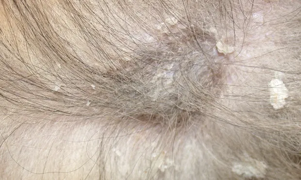
Scale forming an epidermal collarette. Note the circular pattern formed by the lesion.
Gender
Equal incidences
Causes
Causes of scale can be divided into congenital and acquired. Since scaling is a reaction pattern, it is important to understand that anything that affects proliferation, differentiation, or desquamation of the epidermis may produce scale.
Acquired: Due to inflammation, endocrinopathies, nutritional factors, and environmental factors
Congenital: Due to CDA, primary seborrhea, sebaceous adenitis, SCS, and ichthyosis
Risk Factors
Acquired: Low fat diet, frequent bathing, low environmental humidity, allergic skin disease, and endocrinopathies
Congenital: Poor genetics/gene pool
Pathophysiology
Acquired:
Endocrinopathies (hypothyroidism, hyperadrenocorticism)
Cause decrease in protein production (enzymes responsible for normal desquamation), changes in cutaneous fatty acid concentrations (increase in oleic acid; oleic acid is not effective in preventing transepidermal water loss [TEWL], the quantity of water that passes through the epidermis and evaporates into the environment), and decrease in sebaceous gland secretion due to atrophy
Environmental factors
Without adequate water content, enzymes necessary for separation of corneocytes (normal desquamation) will not be produced, leading to scale.
Inflammation
Cytokines are produced when the epidermis is damaged. Cytokines and inflammatory eicosanoids stimulate epidermal proliferation in an effort to remove the noxious insult. Epidermal hyperproliferation leads to defective differentiation of the keratinocytes.
Causes of inflammation include:
Allergic skin disease: Atopy, cutaneous adverse food reaction, flea allergy dermatitis
Infectious: Bacterial, fungal (eg, dermatophytosis or Malassezia), ectoparasites (eg, Demodex or Cheyletiella mites, fleas)
Neoplastic: Epitheliotropic lymphoma
Nutritional factors
Deficiencies in a variety of vitamins, minerals, proteins, or essential fatty acids may cause scaling.
Vitamin A-responsive, zinc-responsive, or fatty acid-responsive dermatoses are not deficiencies, but respond to supplementation.
Congenital:
CDA: Clumping of melanin with the hair shaft results in fragile hairs that fracture easily.
Ichthyosis: Defects in structure or function of intracellular keratinocyte organelles (lamellar granules), enzymes (transglutamase), and cytoskeleton of the keratinocyte. The end result is increase in cohesiveness of stratum corneum cells or in cellular proliferation.
Primary seborrhea: Cellular defect leading to a hyperproliferative epidermis (decrease in epidermal turnover time). Please note that diagnosis of primary seborrhea is a diagnosis of exclusion.
Sebaceous adenitis: Due to destruction of sebaceous glands
SCS: Due to keratin plugging of sebaceous glands
Waxy scale due to seborrhea, resulting in matted hair.
Signs
History
May point you in the direction of the underlying disease.
Allergic skin disease/ectoparasites: Pruritus
CDA: Nonpruritic alopecia in a young dog
Endocrinopathies: Hypothyroidism-lethargy, heat seeking, weight gain; hyperadrenocorticism-polyuria/polydypsia/polyphagia, panting, muscle weakness. Both of these endocrinopathies are nonpruritic unless there is a secondary bacterial or Malassezia infection.
Ichthyosis: Scaling in a young dog
Physical Examination
May also point you in the direction of the underlying disease.
Allergic skin disease/ectoparasites: Evidence of pruritus (alopecia, crusts, erosions, lichenification, excoriations), presence of parasites
CDA: Alopecia in dilute hair areas, presence of comedones, secondary pyoderma (papules and pustules)
Endocrinopathies:
Hypothyroidism: Weight gain, bilateral symmetrical alopecia, bradycardia, hyperpigmentation, myxedema, superficial bacterial folliculitis, Malassezia infection
Hyperadrenocorticism: Panting, muscle weakness, bilateral symmetrical alopecia, hyperpigmentation, pendulous abdomen, hepatomegaly, superficial bacterial folliculitis, Malassezia dermatitis
Sebaceous adenitis:
Standard poodle form: Seen in standard poodles, Akitas, German shepherd dogs. Signs include adherent white scaling, follicular waxy casts (matted hair from the scale), varying degrees of hypotrichosis (including alopecia), dull appearance to the hair coat, pruritus if a secondary pyoderma (papules and pustules) or Malassezia is present, and loss of curls in standard poodles.
Short-coated form: Seen in Vizslas, dachshunds. Dogs present with annular areas of scaling and alopecia most commonly affecting the trunk. At this stage it is not uncommon to mistake this disease for a superficial bacterial folliculitis. However, failure to respond to appropriate antibiotics (eg, cephalosporins, potentiated amoxicillin, potentiated sulfas, etc) rules out superficial bacterial folliculitis.
SCS: Comedone formation on the dorsum
Diagnosis
Definitive Diagnosis (See Suggested reading below for diagnostics used to identify various conditions)
History
Age of onset
Environment
Topical therapies
Degree of pruritus: If present, consider ectoparasites, allergic skin disease, bacterial pyoderma, or Malassezia dermatitis
Presence of constitutional signs (lethargy, polyuria/polydypsia, excessive panting)
Dermatologic exam
Appearance of hair coat
Texture and density (may be dull, dry, thinning, or normal)
Hypotrichosis or alopecia (if present, may be symmetrical, focal, or multifocal)
Presence of ectoparasites
Primary lesions in addition to scale
Alopecia: Posttraumatic, consider hypersensitivities; spontaneous, consider endocrinopathies, follicular dysplasia (eg, CDA), or drugs/medications (eg, glucocorticoids, chemotherapy)
Comedones: Bacterial, fungal, parasitic (Demodex), endocrinopathy (hyperadrenocorticism), primary cornification defects (eg, SCS, acne) or medications/drugs (eg, glucocorticoids)
Crusts: Bacterial, fungal (dermatophytes, Malassezia), autoimmune (pemphigus, vasculitis), ectoparasites
Follicular casts: Vitamin A-responsive dermatosis, primary seborrhea, sebaceous adenitis, or demodicosis
Hyperpigmentation: Endocrinopathies
Macules/patches: Epitheliotropic lymphoma, hypersensitivities or irritants
Nodules/tumors: Epitheliotropic lymphoma, mast cell tumor
Papules/plaques: Bacterial, fungal, neoplastic (epitheliotropic lymphoma); if not follicularly oriented, consider ectoparasites
Pustules: Bacterial, fungal (dermatophytes, Malassezia), autoimmune (pemphigus foliaceus), parasitic (Demodex)
Laboratory findings
Skin scrapings, impression smears, flea combing to identify ectoparasites
Fungal or bacterial culture
Impression smears to identify Malassezia and bacteria
CBC, chemistry profile, and urinalysis may reveal changes that occur with hypothyroidism (elevated cholesterol, mild nonregenerative anemia) or hyperadrenocorticism (mature neutrophilia, eosinopenia, lymphopenia, elevated alkaline phosphatase, hyposthenuria, proteinuria, lower urinary tract infection)
If hypothyroidism or hyperadrenocorticism suspected: Thyroid testing or adrenal function testing (LDDS or ACTH-stimulation test)
Biopsy to diagnose:
CDA
Ichthyosis
SCS
Sebaceous adenitis
Biopsy will identify:
Allergic skin disease, but not the etiology
Endocrinopathy, but not the etiology
Seborrhea, but not differentiate primary versus secondary
Nutritional-responsive dermatosis, but biopsy may not always differentiate these dermatoses from allergic skin disease
Differential Diagnosis
Depends on age of onset, breed, coat color, presence of pruritus or constitutional signs. See Causes under Profile.
Treatment
Inpatient or Outpatient
Outpatient treatment is normally recommended.
Client Education
Most causes of scaling can be treated but not cured. However, scaling caused by nutritional-responsive or environmental causes, cutaneous adverse food reaction, or ectoparasites can be cured while atopic dermatitis can only be controlled. Please note that any dog affected with diseases that have a genetic basis should be neutered.
Medications
Treat the underlying disease.
Therapy for clinical signs-may include:
Shampoos $
Humectants (applied as a final rinse after a bath or misted directly onto the skin from the bottle) $
Calcitriol $$$-$$$$
10 ng/kg Q 24 H
Weekly measurement of electrolytes and parathyroid hormone is recommended when oral formulation is used.
Synthetic retinoids $$$$
Isotretinoin (1-3 mg/kg PO Q 12-24 H)
Acitretin (0.5-1 mg/kg Q 24 H)
Natural vitamin A $$$$$
625-800 IU/kg PO Q 24 H
Oral supplementation with linoleic acid may be beneficial in cases of scaling. Linoleic acid is the most important fatty acid in regard to preventing TEWL and maintaining barrier function. $
Follow-Up
Patient Monitoring
Since the underlying disease causing scaling is frequently not curable (eg, allergies, CDA, sebaceous adenitis) it is important to examine the patient anytime clinical signs (such as pruritus) recur or new lesions appear. If the dog is on synthetic retinoids (which may cause hypercholesterolemia, hypertrigly-cerolemia, hepatopathies, and decreased tear production), a baseline CBC, serum chemistry profile, and Schirmer's tear test should be performed. These tests should be repeated 30 days after therapy begins and then every 6 months. If the lipid profile of a patient becomes elevated, changing to a low fat diet will frequently resolve it.