Canine Protein Losing Enteropathy
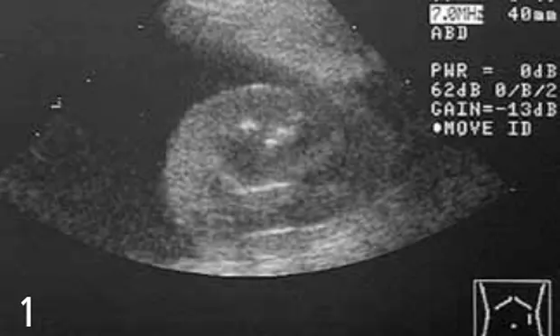
Transverse ultrasonographic image of normal-appearing left kidney surrounded by anechoic ascites. Hyperechoic tissue in the near field is body wall and musculature.
A 4-year-old, spayed female Rottweiler presented with weight loss, diarrhea, and vomiting.
History. The dog had a 1.5-month history of weight loss and small bowel diarrhea characterized as a large volume of semiformed feces several (3 to 4) times a day with no blood or mucus. Decreased appetite and once-a-day vomiting had begun 1 week before presentation. Results of limited blood analysis done 2 weeks before included the following abnormalities:
albumin 0.7 g/dl (reference range, 2.7 to 3.8 g/dl)
ALT 108 U/L (reference range, 10 to 100 U/L)
globulin 1.9 g/dl (reference range, 2.5 to 4.5 g/dl).
The dog was receiving monthly heartworm preventive and had received routine vaccinations 8 months previously.
Physical Examination. Ascites, edema of the distal limbs and ventral neck, and a body condition score of 1.5/5 were found on physical examination; all other physical findings were within normal limits.
Diagnostics. The results of CBC, chemistry panel, urinalysis, fecal analysis, and bile acids test that were done initially are given at left.
Ask yourself ...Which of the following is the most appropriate next step in the diagnostic workup?A. administration of fenbendazoleB. abdominal radiographsC. abdominal ultrasonography followed by endoscopic intestinal biopsiesD. exploratory celiotomyE. none-administer injection of dexamethasone
Correct Answer: CAbdominal ultrasonography followed by endoscopic intestinal biopsies
Panhypoproteinemia most often indicates protein-losing enteropathy. This disorder causes loss of both albumin and globulin through the damaged intestinal wall. However, it is important to rule out other causes of hypoalbuminemia, such as liver dysfunction and protein-losing nephropathy, before invasive diagnostic testing.
Abdominal radiographs may not be helpful. In most animals with clinical signs consistent with gastrointestinal disease, abdominal radiographs are invaluable and should be part of the diagnostic workup. Radiographs may reveal a variety of abnormalities, such as foreign objects, intussusception, masses, and gastric malpositioning. However, in cases of animals with ascites, poor abdominal detail will prevent identification of abdominal structures, so abdominal ultrasonography is more helpful to rule out masses (Figure 1).
Endoscopy may be the preferred method of obtaining biopsies in patients with panhypoproteinemia. Although exploratory celiotomy enables collection of full-thickness biopsies of several areas of the intestinal tract, this procedure may not be optimal in a patient with hypoalbuminemia. Such patients have delayed wound healing and may thus be at greater risk for dehiscence of intestinal biopsy sites.
Further Diagnostics: Endoscopy with histopathologic study of multiple biopsies.

Endoscopy. The esophageal and gastric mucosa appeared normal. The duodenal mucosa appeared mildly granular with many pinpoint white areas and was moderately friable (Figure 2). Several biopsies were submitted for histopathologic evaluation. Although lymphangiectasia can be presumed on the basis of endoscopic appearance, biopsies are still indicated to determine whether the protein-losing enteropathy is a primary or a secondary disorder.
Figure 2. Endoscopy of the proximal duodenum. Note the granular appearance of the mucosa and the multiple white pinpoint foci representing dilated lacteals.
Histopathology. Duodenum-moderate eosinophilic enteritis with dilated lacteals. Therapy should include dietary and medical management. Protein-losing enteropathy (PLE) secondary to inflammation can often be controlled with management of the underlying disease. The prognosis for the PLE may be better if an underlying inflammatory condition is present as compared to primary lymphangiectasia. Until control of the inflammation is achieved, dietary management is very important. Long-chain triglycerides should be avoided in these patients because they are a major stimulus for lymph flow in the intestine. Therefore, low-fat diets have been recommended. Most patients with the disorder are quite thin, have poor body condition, and may not be able to maintain or gain weight on fat- and calorie-restricted diets. Medium-chain triglycerides are thought to be absorbed directly into the portal blood, thus bypassing the lymphatics, and have been recommended as a supplement in patients with abnormal intestinal lymphatics. A combination of diets or a low-fat diet supplemented with medium-chain triglyceride oil may be beneficial in these patients.
Control of inflammation can be achieved with a variety of medications. The most classically used medications are corticosteroids, either alone or in combination with another immunosuppressive drug. This dog was treated with a combination of Hill's R/D and Purina EN formulas, prednisone 2 mg/kg/d initially, and azathioprine 2 mg/kg/d initially. Laboratory values returned to within normal reference range and the edema resolved completely. Once the protein levels have returned to normal and clinical signs have improved or resolved, the medications can be slowly tapered, one at a time. This dog is currently maintained on Purina EN diet and azathioprine 1 mg/kg every other day. Its current body condition is 3.5/5.
At a Glance
Dietary management: Feed a low-fat, highly digestible, medium-chain triglyceride diet.
Medical management: Give corticosteroids alone or combine with another immunosuppressive drug.
Taper medications slowly (20% to 25% every 3 to 4 weeks) once the clinical signs resolve and serum protein levels become normal. Taper medications one at a time down to the lowest effective dose of each. It is possible in some cases to discontinue medications completely, but monitor the patient closely.
Monitor clinical signs and serum protein levels (especially albumin) to gauge response to therapy.
Continue to maintain a highly digestible diet.
Take-Home Messages
In patients with severe panhypoproteinemia, protein-losing enteropathy is likely, even if the patient does not have severe diarrhea.
Patients with severe hypoalbuminemia are at risk for dehiscence of incisions and hypovolemia.
A combination of dietary and medical management is important in the treatment of patients with secondary lymphatic obstruction.
PROTEIN-LOSING ENTEROPATHY • Lisa E. Moore
Suggested Reading
Clinical, clinicopathologic, radiographic, and ultrasonographic characteristics of intestinal lymphangiectasia in dogs: 17 cases (1996-1998). Kull PA, Hess RS, Craig LE, et al. JAVMA 219:197-202, 2001.Idiopathic inflammatory bowel disease in dogs and cats: 84 cases (1987-1990). Jergens AE, Moore FM, Haynes JS, Miles KG. JAVMA 201:1603-1608, 1992.Lymphocytic-plasmacytic enteritis in 24 dogs. Jacobs G, Collins-Kelly L, Lappin M, Tyler D. J Vet Intern Med 4:45-53, 1990.Malabsorption, small intestinal bacterial overgrowth, and protein-losing enteropathy. Williams DA. In Guilford WG, Center SA, Strombeck DR, et al (eds): Strombeck's Small Animal Gastroenterology. Philadelphia: WB Saunders, 1996, pp 375-380.Protein losing enteropathies. Moore LE. In Bonagura JD (ed): Kirk's Current Veterinary Therapy XIII Small Anim Prac. Philadelphia: WB Saunders, 2000, pp 641- 643.